
Lichen Planus: The Itchy Purple Rash That Can Affect Skin, Nails, and Mouth
A rash that is intensely itchy, distinctly purple, and appears simultaneously on the wrists, mouth, and nails presents a clinical picture unlike almost any other skin condition. This distinctive combination points firmly toward lichen planus.
Lichen planus is a chronic inflammatory condition that can affect the skin, mucous membranes, nails, scalp, and genitalia. Its causes involve immune dysregulation, and while it often resolves over time, certain forms persist and cause significant long-term discomfort and potential complications.
Understanding lichen planus causes, recognising its many presentations, and knowing what treatments work helps affected individuals navigate a condition that can be as complex as it is uncomfortable. Early diagnosis prevents unnecessary suffering and serious complications.
What Is Lichen Planus?
Lichen planus is a chronic immune-mediated inflammatory disorder affecting stratified squamous epithelium, which includes the outer skin layer, the lining of the mouth, and the surface of the nails. The name “lichen” refers to the lichen-like appearance of the clustered skin lesions, while “planus” means flat.
The condition affects approximately 0.5 to 2% of the global population, occurring across all ethnicities, ages, and backgrounds. It most commonly develops in adults between 30 and 60 years of age and shows a slight predilection for women, particularly in oral forms.
Cutaneous Versus Mucosal Lichen Planus
Lichen planus manifests in two broad anatomical categories. Cutaneous lichen planus affects the skin, scalp, and nails, while mucosal lichen planus involves the mouth, genitalia, and other mucous membrane surfaces.
Approximately half of people with cutaneous lichen planus also develop oral lesions. Conversely, people with oral lichen planus may have exclusively mucosal disease without visible skin involvement, making awareness of both forms important for comprehensive clinical recognition.
The Risk of Malignant Transformation
Oral lichen planus, particularly the erosive subtype, carries a recognised risk of malignant transformation to oral squamous cell carcinoma. The estimated lifetime transformation risk ranges from 0.5 to 2.5%, making regular specialist monitoring an essential component of long-term oral lichen planus management.
This malignant potential distinguishes oral lichen planus from purely cosmetic skin conditions and justifies ongoing surveillance even during periods when symptoms appear controlled. Any new ulceration, induration, or rapidly changing lesion within oral lichen planus warrants urgent biopsy.
Lichen Planus Causes: The Immune Mechanism
Lichen planus results from a T cell-mediated autoimmune attack on basal keratinocytes, the cells forming the deepest layer of the epithelium. CD8+ cytotoxic T cells recognise altered or foreign antigens expressed on basal keratinocyte surfaces and mount a destructive immune response at the dermoepidermal or epithelial-connective tissue junction.
This immune attack triggers apoptosis, meaning programmed cell death, of basal keratinocytes, producing the characteristic histopathological features of lichen planus. The precise antigen triggering this immune response remains unknown in most idiopathic cases.
Hepatitis C Virus and Lichen Planus
Hepatitis C virus (HCV) infection has the strongest and most consistently demonstrated association with lichen planus among all infectious triggers. Studies from Mediterranean, Asian, and South American populations show significantly higher rates of HCV infection in lichen planus patients compared to controls.
The association is particularly strong for oral and cutaneous forms and may reflect molecular mimicry, where viral antigens resemble self-proteins and trigger cross-reactive immune responses. All patients with lichen planus should undergo HCV serological testing, as successful antiviral treatment can improve lichen planus activity in HCV-positive cases.
Drug-Induced Lichenoid Reactions
Numerous medications can trigger lichenoid drug reactions that closely mimic idiopathic lichen planus both clinically and histologically. These drug-induced forms are called lichenoid reactions rather than true lichen planus, though they are clinically indistinguishable without careful medication history assessment.
Common culprit medications include certain antihypertensives including beta-blockers, angiotensin-converting enzyme inhibitors, and thiazide diuretics, as well as antimalarials, gold compounds, and non-steroidal anti-inflammatory drugs. Drug withdrawal often produces improvement or complete resolution of lichenoid reactions when the causative agent is identified and discontinued.
Other Triggers and Associations
Dental restorative materials, particularly amalgam fillings, can trigger oral lichen planus through a contact hypersensitivity mechanism in susceptible individuals. Replacing amalgam restorations sometimes produces improvement in adjacent oral lichen planus lesions, though this relationship is not consistent across all affected people.
Psychological stress reliably exacerbates lichen planus activity through neuroimmunological pathways and represents one of the most consistently reported aggravating factors by patients. Autoimmune conditions including thyroid disease, primary biliary cholangitis, and inflammatory bowel disease associate with lichen planus at higher rates than the general population.
Recognising the Classic Skin Features of Lichen Planus
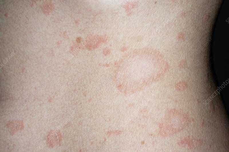
The cutaneous lesions of lichen planus follow a highly characteristic clinical pattern that dermatologists describe using the six Ps: planar, purple, polygonal, pruritic, papules, and plaques. These six descriptors together capture the essence of the classic lichen planus skin lesion.
Each individual lesion is a flat-topped, purple or violaceous, polygonal papule ranging from a few millimetres to over a centimetre in diameter. The flat tops of the lesions often show a fine, lacy white pattern called Wickham striae when examined under dermatoscopy or with a magnifying loupe.
Wickham Striae: A Diagnostic Clue
Wickham striae are fine, white, lacy lines visible on the surface of lichen planus lesions and represent one of the most clinically useful diagnostic features of the condition. They reflect the histopathological changes occurring at the dermoepidermal junction and are most clearly visible when a drop of oil is applied to the lesion surface before examination.
These striae are not visible on all lesions, particularly in very early or very active, eroded lesions. However, their presence on even a proportion of the skin lesions in a widespread rash provides a highly specific diagnostic pointer that guides confident clinical diagnosis.
Body Distribution of Cutaneous Lichen Planus
Cutaneous lichen planus preferentially affects the flexural surfaces of the wrists, forearms, lower legs, and ankles. The wrists represent the single most commonly involved site, and bilateral wrist involvement with typical purple polygonal papules is virtually pathognomonic of lichen planus.
The lower back, sacrum, and shins are also frequently affected. Interestingly, the face is rarely involved in typical cutaneous lichen planus, which helps distinguish it from conditions such as lupus erythematosus and dermatomyositis that commonly affect facial skin.
Koebner Phenomenon in Lichen Planus
Lichen planus demonstrates the Koebner phenomenon, meaning new lesions develop at sites of skin trauma such as scratches, pressure, or surgical wounds. This phenomenon explains why lichen planus lesions sometimes appear in linear arrangements along scratch marks.
Awareness of the Koebner phenomenon guides patient counselling about avoiding skin trauma and tight clothing over affected areas. It also cautions clinicians against unnecessary skin biopsies in multiple sites, as procedural trauma can trigger new lesion formation.
Oral Lichen Planus: Patterns and Complications
Oral lichen planus represents the most clinically significant and potentially dangerous form of the condition due to its malignant transformation risk and significant impact on eating, speaking, and quality of life. It affects approximately 1 to 2% of the global population, making it one of the most common oral mucosal conditions seen in dental and dermatology practice.
Six clinical forms of oral lichen planus exist: reticular, erosive, atrophic, bullous, plaque-like, and papular. The reticular form, characterised by white lacy lines called Wickham striae on the buccal mucosa, is the most common and generally least symptomatic.
Erosive Oral Lichen Planus
Erosive oral lichen planus is the most painful and clinically significant form. It causes raw, irregularly shaped erosions on the tongue, inner cheeks, gums, and palate that make eating, drinking, and speaking extremely painful.
The gingival form of erosive oral lichen planus, called desquamative gingivitis, causes the gum tissue to become bright red, fragile, and prone to bleeding even with gentle toothbrushing. Oral hygiene becomes painful and difficult, compounding gum disease risk and periodontal complications over time.
Monitoring for Oral Cancer Risk
The malignant transformation risk in oral lichen planus demands regular specialist monitoring every six to twelve months depending on subtype and activity level. Erosive forms and those affecting the tongue carry the highest transformation risk and require more frequent surveillance.
Any new hard, fixed, rapidly enlarging, or non-healing area within oral lichen planus should undergo immediate biopsy to exclude squamous cell carcinoma. Patients should understand this monitoring requirement and report any concerning new changes between scheduled appointments without waiting.
Nail Lichen Planus
Lichen planus affects the nails in approximately 10% of people with the condition, and nail involvement can occasionally occur without any skin or mucosal lesions. Nail lichen planus causes a range of changes reflecting the immune attack on the nail matrix and nail bed.
Early nail lichen planus produces longitudinal ridging, thinning, and fragility of the nail plate. These changes may affect one or several nails simultaneously and can be subtle enough to escape notice in their earliest stages.
Severe Nail Involvement and Permanent Damage
Progressive nail lichen planus causes more dramatic changes including onycholysis, meaning separation of the nail from its underlying bed, pterygium formation, and eventually permanent nail loss. Pterygium unguis, a pathognomonic feature of nail lichen planus, describes a triangular scar tissue that grows from the proximal nail fold over the nail surface, permanently obliterating the nail.
Prompt treatment of nail lichen planus during its active phase can prevent permanent nail matrix destruction and preserve nail function. Delayed treatment after pterygium formation and nail destruction has occurred cannot restore the lost nail structure.
Lichen Planopilaris: Scalp Involvement
Lichen planopilaris (LPP) is the follicular variant of lichen planus affecting the scalp hair follicles. It causes a primary scarring alopecia, meaning irreversible hair follicle destruction, that produces permanent patchy hair loss without the possibility of spontaneous regrowth.
Active LPP presents with perifollicular erythema and scaling around individual hair follicles at the periphery of advancing patches. Affected people describe scalp burning, itching, and tenderness, which indicate ongoing active disease requiring systemic treatment.
Frontal Fibrosing Alopecia
Frontal fibrosing alopecia (FFA) is now classified as a variant of lichen planopilaris and represents the most rapidly increasing primary scarring alopecia worldwide. It causes a slowly progressive recession of the frontal and temporal hairline accompanied by loss of eyebrows and occasionally eyelashes.
FFA predominantly affects postmenopausal women, though it increasingly affects younger women and men. The cause of its remarkable global increase remains under active investigation, with sunscreen ingredients, hormonal changes, and dietary factors all proposed as potential contributors.
Diagnosing Lichen Planus
Most lichen planus diagnoses rest on careful clinical assessment of the characteristic lesion morphology, distribution, and history. An experienced dermatologist can diagnose cutaneous lichen planus with confidence in classic presentations without requiring biopsy.
Atypical presentations, mucosal involvement requiring malignancy exclusion, and nail disease where clinical features alone may be insufficient all benefit from biopsy confirmation.
Histopathological Features on Biopsy
A skin or mucosal biopsy of lichen planus shows a distinctive histological pattern comprising dense band-like lymphocytic infiltrate at the dermoepidermal junction, liquefactive degeneration of basal keratinocytes, and colloid bodies representing apoptotic keratinocytes within the dermis.
Saw-tooth rete ridges and hypergranulosis, meaning thickening of the granular layer of the epidermis, further characterise cutaneous lichen planus. These combined histopathological features provide diagnostic confirmation and help exclude lichenoid drug reactions, lupus, and other interface dermatitides.
Baseline Investigations
Hepatitis C serology should be performed in all newly diagnosed lichen planus patients given the established viral association. Liver function tests accompany HCV testing and screen for hepatic involvement that might influence treatment decisions.
Thyroid function testing and autoimmune antibody screening identify associated conditions requiring independent management. In oral lichen planus, direct immunofluorescence of perilesional tissue helps distinguish lichen planus from pemphigoid and other bullous conditions that can produce similar mucosal erosions.
Treating Lichen Planus: A Stepwise Approach
Treatment goals for lichen planus include relieving itching and pain, reducing active inflammation, preventing complications, and monitoring for malignant transformation in oral disease. No treatment cures lichen planus, but effective therapy controls symptoms and reduces disease activity substantially.
Most cutaneous lichen planus resolves spontaneously within one to two years, reducing the need for prolonged systemic therapy in milder cases. Mucosal and nail forms require more sustained treatment due to their greater chronicity and complication risks.
Topical Corticosteroids as First-Line Treatment
Potent topical corticosteroids are the first-line treatment for cutaneous and oral lichen planus. High-potency preparations such as clobetasol propionate applied directly to active lesions reduce inflammation, relieve itching, and accelerate resolution in many cases.
For oral lichen planus, topical steroid preparations including clobetasol gel, triamcinolone acetonide in oral paste, and budesonide mouthwash reduce mucosal inflammation and erosion severity effectively. Application immediately after meals and at bedtime maximises contact time with oral mucosal lesions.
Topical Calcineurin Inhibitors
Topical tacrolimus 0.1% ointment and pimecrolimus 1% cream provide steroid-free anti-inflammatory options for cutaneous and oral lichen planus. These agents are particularly valuable for long-term maintenance therapy in areas where corticosteroid side effects would accumulate with extended use.
Oral tacrolimus has demonstrated efficacy specifically for erosive oral lichen planus in multiple clinical studies. The absence of mucosal atrophy risk with tacrolimus, unlike with prolonged topical corticosteroids, makes it well-suited to oral mucosa management in erosive disease requiring extended treatment.
Systemic Treatments for Severe Lichen Planus
Widespread cutaneous lichen planus, severe erosive oral disease, or nail and scalp lichen planus often requires systemic immunosuppressive therapy. Oral prednisolone at short tapering courses provides rapid symptom control during severe acute flares.
Hydroxychloroquine, acitretin, cyclosporine, and methotrexate all have evidence supporting their use in refractory lichen planus unresponsive to topical treatments. Choosing between these agents depends on the predominant disease location, severity, patient comorbidities, and tolerability preferences.
Emerging Targeted Therapies
Growing understanding of lichen planus immunopathology has identified targeted therapeutic opportunities currently under investigation. JAK inhibitors, which block the interferon-gamma signalling driving T cell-mediated keratinocyte destruction, show promising early results in pilot studies.
Dupilumab has demonstrated benefit in small case series of oral and cutaneous lichen planus, particularly in individuals with concurrent atopic features. These emerging options provide hope for patients with treatment-resistant disease who have exhausted conventional therapeutic approaches.
Frequently Asked Questions About Lichen Planus
What causes lichen planus to develop?
Lichen planus develops through a T cell-mediated immune attack on basal keratinocytes at the skin or mucosal surface. Hepatitis C virus infection is the strongest identified infectious trigger, and all patients should undergo HCV testing at diagnosis. Other triggers include specific medications, dental materials, and psychological stress, though in many cases no specific cause is identified and the condition is classified as idiopathic.
Is oral lichen planus dangerous?
Oral lichen planus is not immediately dangerous but carries a recognised risk of malignant transformation to oral squamous cell carcinoma, estimated at 0.5 to 2.5% over a lifetime. Erosive forms and tongue involvement carry the highest transformation risk and require regular specialist monitoring every six to twelve months. Reporting any new, hard, or rapidly changing area within oral lichen planus lesions to a specialist immediately is important for early cancer detection.
Does lichen planus go away permanently?
Cutaneous lichen planus typically resolves spontaneously within one to two years in most cases, though post-inflammatory pigment changes may persist for months afterwards. Oral lichen planus tends to be more chronic and persistent, often requiring ongoing maintenance treatment for years. Nail and scalp lichen planus can cause permanent structural damage if not treated promptly, making early intervention essential for preserving nail and hair follicle integrity.
How is lichen planus different from psoriasis?
Lichen planus and psoriasis both cause chronic inflammatory skin plaques but are entirely distinct conditions with different mechanisms, appearances, and treatments. Lichen planus produces flat-topped, purple, polygonal papules primarily on the wrists and flexural surfaces with characteristic Wickham striae. Psoriasis produces thicker, silvery-scaled, salmon-pink plaques predominantly on extensor surfaces and the scalp, often associated with nail pitting and arthritis rather than the nail changes typical of lichen planus.
Can lichen planus affect the genitalia?
Yes, lichen planus commonly affects the genitalia in both men and women, often producing painful erosive lesions similar to those seen in oral lichen planus. Genital lichen planus in women can cause vulval and vaginal erosions that make sexual activity, urination, and daily comfort extremely difficult. In men, the glans penis and foreskin are most commonly affected. Genital lichen planus requires specialist management and monitoring due to its chronic course, painful nature, and potential complications including scarring and stenosis.
Managing Lichen Planus With Knowledge, Vigilance, and Specialist Care
Lichen planus is a condition of remarkable clinical diversity, ranging from a self-limiting purple wrist rash to a chronic, painful, and potentially premalignant oral disease. Its many faces make accurate diagnosis essential, and its complications make specialist care genuinely important.
Modern dermatology and oral medicine offer an expanding range of effective treatments that bring meaningful relief to people living with active lichen planus. From first-line topical corticosteroids and calcineurin inhibitors to emerging JAK inhibitors and biological agents, the therapeutic landscape continues to broaden.
Above all, understanding the malignant transformation risk in oral lichen planus, the irreversibility of nail and scalp damage without prompt treatment, and the importance of hepatitis C screening transforms this manageable skin condition from a source of anxiety into a condition where informed, proactive engagement genuinely changes long-term outcomes.
Disclaimer:
This article is intended for general informational purposes only. It does not constitute medical advice, diagnosis, or treatment. Always consult a qualified healthcare professional for any medical concerns.
References:
- Lichens are fascinating organisms formed through the symbiotic relationship between fungi and photobionts, which are typically green algae or cyanobacteria.
- The vulva comprises external female genitalia including: mons pubis (hair-bearing fatty tissue over pubic bone
- A quirky new beauty trend is taking social media by storm, and it’s all about the color green! Dubbed the “green nail theory
Observer Voice is the one stop site for National, International news, Sports, Editor’s Choice, Art/culture contents, Quotes and much more. We also cover historical contents. Historical contents includes World History, Indian History, and what happened today. The website also covers Entertainment across the India and World.