
Bullous Pemphigoid: The Blistering Skin Condition Most Common in Older Adults
Most people associate blistering skin conditions with burns, allergic reactions, or infections. Bullous pemphigoid presents a very different and more complex clinical picture.
This chronic autoimmune skin disease causes large, tense blisters to form across the body, predominantly affecting people over 70 years of age. The immune system mistakenly attacks proteins in the skin, triggering intense inflammation and blister formation that can become life-threatening without treatment.
Bullous pemphigoid causes significant morbidity in older adults and carries a substantial one-year mortality rate, partly due to disease severity and partly due to the serious side effects of treatment. Early diagnosis and expert management save lives and preserve quality of life.
What Is Bullous Pemphigoid?
Bullous pemphigoid is a chronic autoimmune blistering disease affecting the skin and, less commonly, the mucous membranes. The term “bullous” refers to large blisters called bullae, while “pemphigoid” indicates a resemblance to pemphigus without sharing its exact mechanism.
Unlike pemphigus vulgaris, which attacks proteins deep within the epidermis, bullous pemphigoid targets proteins at the junction between the epidermis and the dermis beneath it. This deeper attack site produces blisters that are tighter, more resilient, and less prone to spontaneous rupture.
How Bullous Pemphigoid Differs From Other Blistering Diseases
The anatomical location of blistering determines much of the clinical behaviour seen in bullous pemphigoid. Sub-epidermal blisters, forming beneath the skin’s outermost layer, are substantially more robust than the intra-epidermal blisters characteristic of pemphigus vulgaris.
This difference explains why bullous pemphigoid blisters remain intact longer before rupturing. However, when blisters do rupture or when eosinophilic inflammation dominates the clinical picture, significant skin damage and secondary infection risk still occur.
Why Bullous Pemphigoid Predominantly Affects Older Adults
Bullous pemphigoid is strongly age-associated, with incidence rising sharply in people over 70 years old. The annual incidence in people over 80 is approximately 150 to 300 cases per million, making it the most common autoimmune blistering disease in older populations worldwide.
Age-related immune system changes, including immunosenescence and increased autoreactive antibody production, likely contribute to this strong age association. The condition is rare in younger adults and exceptional in children.
The Autoimmune Mechanism Behind Bullous Pemphigoid
Bullous pemphigoid develops when the immune system produces autoantibodies targeting two structural proteins at the dermoepidermal junction. These proteins, BP180 (also called type XVII collagen) and BP230, form part of specialised adhesion structures called hemidesmosomes.
Hemidesmosomes anchor the epidermis to the deeper dermis, providing essential structural integrity to the entire skin surface. When autoantibodies attack BP180 and BP230, these anchoring structures lose their function and the skin layers separate, forming blisters.
BP180: The Primary Autoantigen
BP180 is the primary target autoantigen in bullous pemphigoid and the most clinically important. Antibodies against BP180 activate complement proteins and recruit eosinophils, a type of immune cell, to the affected skin area.
Eosinophils release destructive enzymes that degrade the dermoepidermal junction proteins directly, amplifying tissue damage beyond what antibody binding alone causes. The resulting inflammation, fluid accumulation, and structural separation produce the characteristic clinical features of this condition.
BP230 and Its Role in Disease
BP230 is an intracellular protein forming part of the hemidesmosomal plaque. Anti-BP230 antibodies correlate less strongly with disease activity than anti-BP180 antibodies but contribute to the overall pathological process.
Measuring both anti-BP180 and anti-BP230 antibody levels in the blood helps confirm diagnosis and monitor disease activity over time. Serial antibody level measurements guide treatment decisions more reliably than clinical assessment alone in many cases.
What Triggers the Autoimmune Response?
In most cases, the precise trigger initiating the autoimmune response in bullous pemphigoid remains unclear. However, several factors associate with disease onset in susceptible older individuals.
Medications including diuretics such as furosemide, gliptins used in diabetes management, and certain antibiotics can trigger drug-induced bullous pemphigoid. Neurological conditions including multiple sclerosis, Parkinson’s disease, dementia, and stroke also associate strongly with bullous pemphigoid development, suggesting shared pathogenic mechanisms.
Bullous Pemphigoid Causes and Risk Factors
Understanding the risk factors for bullous pemphigoid helps clinicians identify susceptible patients and initiate prompt investigation when characteristic symptoms emerge. Several independent factors raise disease risk significantly beyond age alone.
Neurological disease represents the strongest non-age-related risk factor for bullous pemphigoid. Studies consistently demonstrate two to three-fold elevated risk in people with Parkinson’s disease, dementia, stroke, and epilepsy compared to neurologically healthy older adults.
Medication-Induced Bullous Pemphigoid
Drug-induced bullous pemphigoid has increased considerably in recent years alongside expanded use of the implicated medication classes. Dipeptidyl peptidase-4 (DPP-4) inhibitors, commonly called gliptins, used widely in type 2 diabetes management, associate strongly with bullous pemphigoid development.
Unlike spontaneous bullous pemphigoid, drug-induced cases often show less mucosal involvement and more atypical clinical features. Identifying and withdrawing the culprit medication forms an essential early step in managing drug-induced cases, though immunosuppressive treatment usually remains necessary.
Other Contributing Risk Factors
Physical trauma to the skin, ultraviolet radiation exposure, and certain vaccinations have been reported as potential bullous pemphigoid triggers in individual cases. These triggers likely unmask pre-existing autoimmune susceptibility rather than independently causing the condition.
Genetic factors also play a role, with certain HLA alleles associating with increased disease susceptibility. However, bullous pemphigoid does not follow clear familial inheritance patterns, distinguishing it from some other autoimmune conditions with stronger genetic transmission.
Recognising the Symptoms of Bullous Pemphigoid
Bullous pemphigoid typically develops in two distinct clinical phases before the full blistering picture emerges. Recognising the pre-bullous phase allows earlier diagnosis and treatment before widespread blistering develops.
Intense generalised itching, often without any visible skin changes, characterises the earliest phase of many bullous pemphigoid cases. This pruritus can precede visible blistering by weeks to months and is frequently misattributed to other causes in older adults.
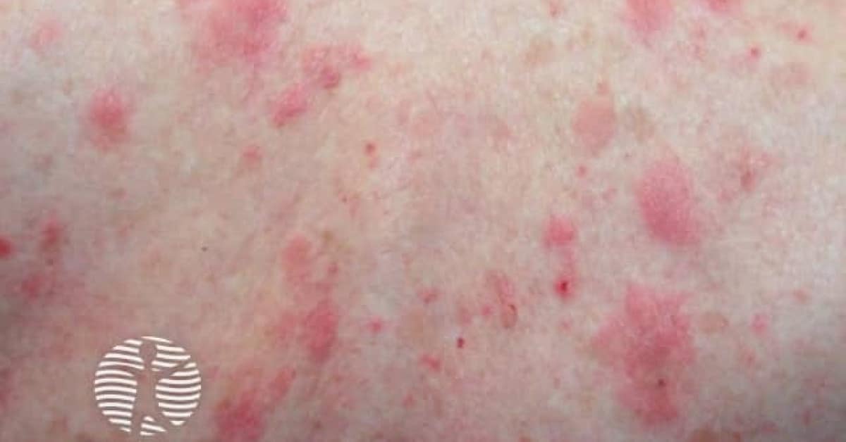
Pre-Bullous Phase: Itching and Eczema-Like Changes
The pre-bullous phase of bullous pemphigoid presents with severe itching accompanied by eczema-like red patches, hives, or thickened plaques on the skin. These changes lack the diagnostic specificity of blisters and frequently lead to delayed or incorrect diagnosis.
Clinicians evaluating older adults with unexplained severe pruritus and eczematous skin changes should maintain a high index of suspicion for bullous pemphigoid. Blood tests measuring eosinophil counts and anti-BP180 antibody levels can confirm the diagnosis before blisters appear.
The Bullous Phase: Large Tense Blisters
The bullous phase brings the appearance of large, tense, fluid-filled blisters arising on inflamed red skin or occasionally on apparently normal skin. These blisters range from one to several centimetres in diameter and feel firm and resilient compared to the fragile blisters of pemphigus vulgaris.
Blisters form most commonly on the flexural surfaces including the inner thighs, forearms, axillae, and abdomen. The fluid inside blisters is usually clear but may become blood-tinged, particularly in more inflamed or traumatised areas.
Distribution Patterns and Affected Areas
Bullous pemphigoid blisters tend to favour the lower trunk, inner thighs, and flexural skin creases. The face and scalp are less commonly involved compared to pemphigus vulgaris, though these areas can develop lesions in severe cases.
Mucous membrane involvement occurs in approximately 10 to 30% of bullous pemphigoid cases, far less frequently than in pemphigus vulgaris. When mucosal lesions do develop, they affect the mouth most commonly, causing painful erosions that complicate eating and oral hygiene.
Complications From Ruptured Blisters
When blisters rupture, they leave behind moist, painful erosions that take considerable time to heal in older adults with compromised skin repair capacity. Secondary bacterial infection of these open wounds is common and significantly worsens outcomes.
Cellulitis, septicaemia, and wound complications from ruptured bullous pemphigoid blisters contribute meaningfully to the elevated one-year mortality seen in affected older adults. Prompt wound management alongside specific immunosuppressive treatment reduces these preventable deaths.
Diagnosing Bullous Pemphigoid
Diagnosing bullous pemphigoid requires combining clinical assessment with specific laboratory investigations. Several other skin conditions, including eczema, drug reactions, linear IgA disease, and pemphigus vulgaris, can produce similar clinical appearances requiring differentiation.
A skin biopsy from the edge of an intact blister provides histopathological evidence showing sub-epidermal blister formation with a prominent eosinophil-rich inflammatory infiltrate. These characteristic findings strongly suggest bullous pemphigoid and direct further confirmatory immunological testing.
Direct Immunofluorescence Testing
Direct immunofluorescence (DIF) on perilesional normal skin is the gold standard investigation for confirming bullous pemphigoid diagnosis. Fluorescent-labelled antibodies detect linear deposits of IgG and complement C3 along the dermoepidermal junction.
This characteristic linear fluorescence pattern directly reflects the pathological process of autoantibody attack at the hemidesmosomal attachment site. A positive DIF result with the appropriate clinical and histological context confirms the bullous pemphigoid diagnosis with high confidence.
Serological Tests and Antibody Measurement
Blood tests measuring circulating anti-BP180 and anti-BP230 IgG antibody levels confirm the diagnosis serologically. Anti-BP180 ELISA demonstrates high sensitivity and specificity for bullous pemphigoid and is widely available in most specialist dermatology centres.
Quantitative anti-BP180 levels correlate with disease activity and treatment response over time. Doctors use serial measurements to guide immunosuppressive dose adjustments, as persistently elevated levels indicate ongoing disease activity even when the skin appears clinically improved.
Excluding Drug-Induced Causes
A thorough medication review forms an essential component of every bullous pemphigoid diagnostic workup. Identifying a temporally related culprit medication, particularly a gliptin or diuretic, changes initial management by prioritising drug withdrawal alongside standard treatment.
Drug-induced bullous pemphigoid may resolve after medication withdrawal alone in some patients, though most still require a course of immunosuppressive therapy. Communicating the suspected drug trigger to the prescribing physician ensures appropriate alternative medications are selected going forward.
Treating Bullous Pemphigoid: A Stepwise Approach
The goals of bullous pemphigoid treatment are to stop new blister formation, heal existing skin erosions, relieve itching, and prevent relapse. Treatment choice depends on disease extent, severity, patient age, comorbid conditions, and ability to tolerate systemic medications.
Most older adults with bullous pemphigoid have multiple medical comorbidities that influence treatment safety and tolerability. Individualised treatment planning by a specialist dermatologist, often in collaboration with geriatric medicine teams, achieves the best clinical outcomes.
Potent Topical Corticosteroids for Localised Disease
High-potency topical corticosteroid creams, particularly clobetasol propionate 0.05%, are the preferred first-line treatment for extensive but non-severe bullous pemphigoid. A landmark French trial demonstrated that topical clobetasol achieved equivalent or superior disease control to oral prednisolone with a lower rate of serious adverse effects.
Applying topical corticosteroids to the entire body surface twice daily during the active phase requires considerable effort and caregiver support in older adults. The treatment burden is significant, but the avoidance of systemic steroid side effects justifies this approach in eligible patients.
Oral Corticosteroids: Benefits and Risks
Oral prednisolone at doses of 0.5 mg per kilogram per day achieves rapid suppression of bullous pemphigoid activity in most patients. This systemic approach is practical for widespread or rapidly progressive disease where topical treatment alone proves insufficient.
However, systemic corticosteroids carry substantially elevated risks in older adults. Infections, osteoporotic fractures, hyperglycaemia, delirium, and adrenal suppression all occur more frequently and severely in this age group, demanding cautious dose escalation and proactive complication prevention.
Steroid-Sparing Agents
Doxycycline, a tetracycline antibiotic, exerts anti-inflammatory effects beyond its antimicrobial properties and has demonstrated modest efficacy in mild to moderate bullous pemphigoid. It offers a valuable treatment option for patients who cannot safely tolerate corticosteroids due to significant medical comorbidities.
Azathioprine, mycophenolate mofetil, and methotrexate serve as steroid-sparing immunosuppressants that allow corticosteroid doses to reduce while maintaining disease control. These agents are introduced after initial disease stabilisation and continued during the slow tapering phase of treatment.
Emerging and Biological Treatments for Bullous Pemphigoid
The expanding understanding of bullous pemphigoid immunopathology has identified several targeted treatment approaches now entering clinical practice. These biological therapies offer hope for patients with refractory disease or those unable to tolerate conventional immunosuppression.
Dupilumab, which blocks IL-4 and IL-13 signalling, has shown promising efficacy in bullous pemphigoid given the prominent type 2 immune response driving disease activity. Several clinical trials are evaluating dupilumab specifically for this indication.
Omalizumab and IgE-Targeting Approaches
Elevated IgE levels and IgE anti-BP180 antibodies feature prominently in bullous pemphigoid pathology. Omalizumab, an anti-IgE monoclonal antibody used in severe asthma, has demonstrated beneficial effects in case reports and small series of bullous pemphigoid patients.
Larger controlled trials are necessary before omalizumab becomes a standard treatment option. Nevertheless, its mechanism directly targets a relevant pathological pathway, making it a rational therapeutic candidate for selected patients with elevated IgE-mediated disease activity.
Rituximab for Refractory Cases
Rituximab depletes B cells responsible for producing the pathogenic anti-BP180 and anti-BP230 antibodies. Case series and small controlled studies demonstrate meaningful responses in refractory bullous pemphigoid that has failed conventional immunosuppressive treatment.
Rituximab use in older adults requires careful benefit-risk assessment given the infection risks associated with B cell depletion in an already immunosenescent population. Specialist centres increasingly consider rituximab for selected patients when standard treatments prove inadequate.
Long-Term Management and Monitoring
Bullous pemphigoid requires long-term clinical follow-up to monitor for relapse, manage treatment side effects, and adjust therapy as disease activity changes. Many patients achieve remission and eventually discontinue treatment, while others require ongoing low-dose maintenance therapy indefinitely.
Anti-BP180 antibody levels guide treatment tapering decisions alongside clinical assessment of blister activity. Persistently elevated antibody levels despite apparent clinical control indicate an ongoing autoimmune process requiring continued treatment.
Predicting and Managing Relapse
Relapse occurs in approximately 30 to 50% of bullous pemphigoid patients after initial remission, most commonly within the first year after treatment discontinuation. Rising anti-BP180 antibody levels during monitoring may predict clinical relapse before blisters reappear.
Prompt treatment reinstitution at the first signs of relapse prevents widespread blister development and its associated complications. Patients and caregivers should receive clear instructions about recognising early relapse signs and contacting their dermatology team without delay.
Mortality Risk in Bullous Pemphigoid
One-year mortality rates for bullous pemphigoid in older adults range from 20 to 40% across published studies. This elevated mortality reflects a combination of disease severity, treatment complications, frailty, and the multiple comorbidities present in affected older individuals.
Aggressive infection prevention, careful corticosteroid dose management, bone protection with calcium and vitamin D supplementation, and proactive management of comorbidities all reduce treatment-related mortality meaningfully. Integrated geriatric and dermatological care achieves the best outcomes in this vulnerable population.
Bullous Pemphigoid and Neurological Disease
The association between bullous pemphigoid and neurological conditions represents one of the most intriguing and clinically important aspects of this disease. Multiple studies confirm two to three-fold elevated bullous pemphigoid risk in people with Parkinson’s disease, Alzheimer’s disease, stroke, and multiple sclerosis.
One proposed mechanism suggests that BP180, which is expressed in both skin and neurological tissue, becomes an antigenic target when neural tissue damage exposes the protein to the immune system. The resulting systemic autoimmune response then cross-reacts with skin BP180 to trigger bullous pemphigoid.
Clinical Implications of the Neurological Link
Clinicians managing older adults with neurological disease should maintain awareness of bullous pemphigoid as a potential complication. New onset of severe pruritus or unexplained skin blisters in patients with Parkinson’s disease or dementia warrants urgent dermatological evaluation.
Conversely, dermatologists diagnosing bullous pemphigoid in a patient without known neurological disease should consider screening for underlying neurological conditions. This bidirectional awareness improves outcomes by facilitating earlier diagnosis and management of both conditions simultaneously.
Managing Bullous Pemphigoid in People With Dementia
People with dementia present particular management challenges in bullous pemphigoid care. Impaired communication makes symptom reporting difficult, topical treatment application requires caregiver involvement, and systemic treatment risks are amplified by cognitive impairment and associated functional decline.
Simplified treatment regimens, close caregiver education, and regular nursing review improve treatment adherence and outcomes in this vulnerable subgroup. Care home staff and community nurses play crucial roles in delivering and monitoring bullous pemphigoid treatment in these patients.
Frequently Asked Questions About Bullous Pemphigoid
What causes bullous pemphigoid to develop?
Bullous pemphigoid causes involve autoimmune production of IgG antibodies targeting BP180 and BP230 proteins at the dermoepidermal junction, triggering blister formation through complement activation and eosinophil recruitment. Age-related immune changes, neurological conditions, and certain medications including gliptins and diuretics increase individual susceptibility significantly. The precise trigger initiating the autoimmune response remains unclear in most cases, though drug withdrawal can resolve disease in medication-induced bullous pemphigoid.
Is bullous pemphigoid life-threatening?
Bullous pemphigoid carries significant mortality risk, particularly in older adults with multiple comorbidities. Published studies report one-year mortality rates of 20 to 40%, reflecting both the dangers of widespread blistering and the serious side effects of immunosuppressive treatment in frail older patients. Prompt specialist diagnosis, careful treatment selection, aggressive infection prevention, and integrated geriatric care substantially improve survival outcomes.
How does bullous pemphigoid differ from pemphigus vulgaris?
Both conditions are autoimmune blistering diseases, but they differ fundamentally in mechanism, clinical features, and severity. Bullous pemphigoid attacks proteins at the dermoepidermal junction, producing tense, resilient blisters that are predominantly a disease of older adults. Pemphigus vulgaris attacks proteins within the epidermis itself, causing fragile blisters and extensive oral mucosal involvement more commonly in middle-aged adults, and historically carried even higher untreated mortality.
Can bullous pemphigoid be cured permanently?
Some patients achieve complete remission and successfully discontinue all treatment, particularly those with mild disease or drug-induced cases where removing the culprit medication resolves the condition. However, relapse occurs in approximately 30 to 50% of patients after treatment withdrawal. Long-term dermatological monitoring remains necessary, and some patients require ongoing low-dose maintenance therapy indefinitely to prevent blister recurrence.
What role does diet or lifestyle play in managing bullous pemphigoid?
No specific diet is proven to treat or prevent bullous pemphigoid, but maintaining good overall nutrition supports skin healing and immune function in affected older adults. Patients on long-term corticosteroids should take calcium and vitamin D supplementation to reduce osteoporosis risk and follow bone-protective dietary guidance. Avoiding identified trigger medications, maintaining skin hygiene, protecting skin from trauma, and adhering consistently to prescribed treatment regimens are the most important practical management steps for affected individuals.
Acting Early Against Bullous Pemphigoid Protects Older Adults
Bullous pemphigoid stands as the most common autoimmune blistering disease in older adults worldwide, yet it remains underrecognised and sometimes mismanaged in the community. Its strong associations with neurological disease and medication use make clinical awareness among all healthcare professionals genuinely essential.
Modern treatment approaches, from potent topical corticosteroids to promising biological therapies, have meaningfully improved outcomes for affected patients. Alongside medical advances, integrated geriatric and dermatological care, caregiver education, and careful complication monitoring all contribute to better survival and quality of life.
Most importantly, recognising the early warning signs of bullous pemphigoid — severe unexplained itching and eczematous skin changes in an older adult — allows diagnosis before widespread blistering establishes itself. Earlier diagnosis means earlier treatment, and earlier treatment saves lives, preserves skin integrity, and protects the independence and dignity that every older person deserves.
Disclaimer:
This article is intended for general informational purposes only. It does not constitute medical advice, diagnosis, or treatment. Always consult a qualified healthcare professional for any medical concerns.
References:
- Pemphigus vulgaris is a chronic autoimmune blistering disorder affecting the skin and mucous membranes.
- Health research encompasses diverse approaches addressing different questions.
- Autoimmune hepatitis is a chronic autoimmune disease where the body’s immune system mistakenly attacks liver cells causing progressive inflammation and scarring.
Observer Voice is the one stop site for National, International news, Sports, Editor’s Choice, Art/culture contents, Quotes and much more. We also cover historical contents. Historical contents includes World History, Indian History, and what happened today. The website also covers Entertainment across the India and World.