
Essential Thrombocythemia: Excess Platelets and Stroke Risk
Most people associate strokes and dangerous blood clots with conditions like high blood pressure, atrial fibrillation, or blocked arteries. Few expect the culprit to be an excess of the very cells the body uses to stop bleeding. Yet for people with essential thrombocythemia, an enormous surplus of platelets — the tiny blood fragments designed to form clots at sites of injury — creates a paradoxical and persistent risk of dangerous clotting throughout the body.
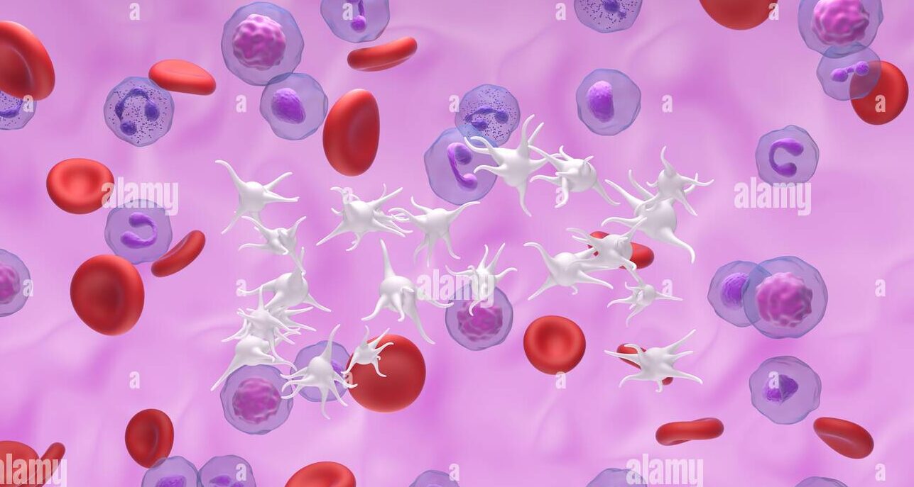
Essential thrombocythemia is a chronic bone marrow condition in which the marrow overproduces platelets far beyond the body’s needs. Instead of the normal platelet count of 150,000 to 400,000 per microlitre of blood, people with ET may have counts of one million or more. These excess platelets are not only numerous — they are functionally abnormal. They activate spontaneously, aggregate inappropriately, and generate blood clots that can block arteries supplying the brain, heart, and limbs.
Essential thrombocythemia excess platelets stroke risk is a connection that surprises many patients at diagnosis — because the condition often produces no symptoms for years before a thrombotic event reveals its presence. Furthermore, ET predominantly affects people in middle age and older adulthood — a population already carrying cardiovascular risk factors that amplify the platelet-driven clotting danger. Consequently, early detection, accurate risk stratification, and consistent preventive treatment are the pillars of managing this condition before catastrophe strikes.
What Is Essential Thrombocythemia?
A Myeloproliferative Neoplasm
Essential thrombocythemia belongs to the same family of bone marrow cancers as polycythemia vera and primary myelofibrosis — called myeloproliferative neoplasms, or MPNs. All three conditions share the fundamental biology of a clonally expanded, genetically mutant haematopoietic stem cell driving excessive blood cell production. In polycythemia vera, red cells predominate. In essential thrombocythemia, platelets predominate. However, the underlying molecular mechanisms overlap significantly. For context on the broader MPN spectrum, see our article on polycythemia vera — too many red blood cells and the clotting risks that follow.
The word essential in essential thrombocythemia means primary — arising from an intrinsic bone marrow disorder rather than as a reactive response to another condition. This distinction is critical because reactive thrombocytosis — elevated platelets responding to infection, inflammation, iron deficiency, or tissue injury — is far more common than ET and carries no significant thrombotic risk. Consequently, distinguishing ET from reactive thrombocytosis is the primary diagnostic challenge when a high platelet count is first discovered.
The Biology of Platelet Overproduction
In healthy bone marrow, megakaryocytes — the large precursor cells that produce platelets by shedding cytoplasmic fragments — respond to a hormone called thrombopoietin. Thrombopoietin signals megakaryocytes to produce more or fewer platelets according to the body’s current needs. When platelet counts are adequate, thrombopoietin signalling dampens megakaryocyte activity. This feedback loop maintains platelet counts within the normal range throughout life.
In essential thrombocythemia, mutations in the genes encoding components of the thrombopoietin signalling pathway — principally JAK2, CALR, and MPL — disrupt this feedback mechanism. The megakaryocyte clone carrying the mutation produces platelets continuously and excessively — independent of whether the body actually needs more platelets. Furthermore, the platelets produced by this abnormal clone are structurally and functionally abnormal — larger than normal, more prone to spontaneous activation, and more likely to aggregate inappropriately. Consequently, the combination of excessive numbers and abnormal function creates a uniquely dangerous pro-thrombotic environment.
The Genetic Drivers of Essential Thrombocythemia
JAK2 V617F Mutation
The JAK2 V617F mutation — the same point mutation that drives polycythemia vera — is present in roughly 50 to 60% of ET patients. In ET, JAK2 V617F produces a less pronounced activation of the JAK-STAT signalling pathway than in PV — explaining why red cell overproduction does not dominate as it does in PV, while platelet overproduction still occurs. Furthermore, JAK2 V617F-positive ET patients tend to have higher haematocrit and white cell counts than their JAK2-negative counterparts — reflecting the broader haematopoietic stimulation that the mutation produces across multiple cell lineages.
JAK2 V617F-positive ET patients also carry a higher thrombotic risk than JAK2-negative patients — making JAK2 mutation status an important component of individual risk assessment at diagnosis. Consequently, JAK2 testing is mandatory in all patients with suspected ET for both diagnostic and prognostic purposes.
CALR Mutations
Calreticulin — encoded by the CALR gene — is a protein normally located in the endoplasmic reticulum of cells. Mutations in CALR — classified as type 1 and type 2 insertions and deletions — are found in roughly 25 to 35% of ET patients. These mutations produce an abnormal calreticulin protein that aberrantly activates the thrombopoietin receptor MPL — driving megakaryocyte proliferation and excess platelet production through a mechanism distinct from JAK2 activation.
CALR-mutated ET patients generally have higher platelet counts but lower thrombotic risk than JAK2-mutated patients. Furthermore, CALR type 1 mutations carry a higher risk of transformation to myelofibrosis than type 2 mutations or JAK2 mutations. Consequently, CALR mutation type provides important prognostic information that refines risk assessment beyond a simple positive or negative result.
MPL Mutations and Triple-Negative ET
Mutations in MPL — the gene encoding the thrombopoietin receptor — are found in roughly 3 to 5% of ET patients. These mutations constitutively activate the receptor — producing the same uncontrolled megakaryocyte stimulation as JAK2 and CALR mutations through a third distinct molecular mechanism. MPL-mutated ET patients show clinical features similar to CALR-mutated ET with high platelet counts and lower thrombotic risk.
Approximately 10 to 15% of ET patients carry none of the three canonical mutations — JAK2, CALR, or MPL. This group — called triple-negative ET — presents diagnostic challenges because the absence of a defining mutation requires particularly careful exclusion of reactive thrombocytosis and other myeloid neoplasms. Furthermore, triple-negative patients generally carry the lowest thrombotic risk among ET subgroups. Consequently, molecular mutation analysis is not merely diagnostic but central to risk stratification and treatment planning in ET.
Symptoms of Essential Thrombocythemia
Thrombotic Events
Essential thrombocythemia excess platelets stroke risk produces its most dangerous consequences through thrombosis — blood clot formation in arteries and veins. Arterial thrombosis dominates in ET — producing stroke, transient ischaemic attack, myocardial infarction, and peripheral arterial occlusion. Stroke and TIA are particularly characteristic of ET — occurring more frequently in younger ET patients than most physicians expect given the benign appearance of the blood count at first glance.
The mechanism of arterial thrombosis in ET involves spontaneous platelet activation and aggregation in small arteries. Activated platelets release thromboxane A2 — a potent vasoconstrictor and platelet activator — that amplifies the clotting response locally. Furthermore, abnormal platelet-endothelial interactions generate microthrombi — tiny clots — in the microcirculation supplying the brain and fingers. Consequently, ET patients may experience recurring transient neurological symptoms — transient vision loss, tingling of fingers, speech difficulty — that reflect microthrombus formation in cerebral and digital vessels.
Erythromelalgia
Erythromelalgia is a distinctive and highly characteristic symptom of essential thrombocythemia. It manifests as episodes of intense burning pain, redness, and warmth in the hands and feet — typically triggered by heat, exercise, or dependent positioning. The episodes last minutes to hours and respond dramatically to low-dose aspirin — a response so characteristic that it provides both diagnostic and therapeutic information simultaneously.
The mechanism involves platelet-mediated microvascular occlusion in the small arteries of the digits. Activated platelets aggregate in these tiny vessels, reducing blood flow and triggering ischaemic pain. Furthermore, the inflammatory mediators released by activated platelets produce the characteristic warmth and redness that accompany the pain. Consequently, erythromelalgia — when present — is almost pathognomonic for ET or polycythemia vera and should immediately prompt platelet count measurement and haematology referral.
Paradoxical Bleeding
Paradoxically, extreme thrombocytosis — platelet counts above 1.5 million per microlitre — can produce bleeding rather than clotting. At very high platelet concentrations, von Willebrand factor — a large protein essential for platelet adhesion to damaged vessel walls — is adsorbed onto the surface of the excess platelets and removed from circulation. This acquired von Willebrand syndrome reduces the effective concentration of functional von Willebrand factor below the level needed for normal haemostasis.
Consequently, patients with extremely high platelet counts may paradoxically experience mucosal bleeding — including nosebleeds, gum bleeding, and gastrointestinal bleeding — despite having an apparently pro-thrombotic high platelet count. Furthermore, this acquired von Willebrand syndrome means that aspirin — by additionally impairing platelet function — can significantly worsen bleeding in patients with platelet counts above 1.5 million. Consequently, aspirin should be withheld in ET patients with platelet counts above this threshold until cytoreductive therapy has reduced the count to safer levels.
How Doctors Diagnose Essential Thrombocythemia
Blood Count and Film
Diagnosing essential thrombocythemia excess platelets stroke risk begins with a full blood count showing a sustained platelet count above 450,000 per microlitre — the lower diagnostic threshold set by the WHO 2016 criteria. Furthermore, counts are typically much higher in ET — often exceeding 600,000 to 1,000,000 per microlitre at diagnosis. The red cell and white cell counts may be mildly elevated — particularly in JAK2-positive cases — but do not meet criteria for PV.
Blood film examination reveals the platelet abnormalities characteristic of ET. Platelets appear large — called giant platelets — and are present in greatly increased numbers. Clumps of platelets may be visible across the film. Furthermore, morphologically dysplastic red cells and white cells — prominent in MDS — are absent in ET. Consequently, the combination of marked thrombocytosis with large but otherwise morphologically normal cells on film points toward a myeloproliferative rather than myelodysplastic process.
Molecular Testing and Bone Marrow Biopsy
Sequential molecular testing — starting with JAK2 V617F, then CALR, then MPL — identifies the driver mutation in roughly 85 to 90% of ET cases. A positive result confirms the clonal nature of the thrombocytosis and supports ET diagnosis. Furthermore, mutation identification guides risk stratification and may influence treatment choices — particularly regarding thrombotic risk and transformation risk differences between mutation subtypes.
Bone marrow biopsy is required for definitive WHO-compliant ET diagnosis. The characteristic finding is a hypercellular marrow with markedly increased numbers of large, mature megakaryocytes showing characteristic staghorn-shaped nuclei — the distinctive nuclear shape of ET megakaryocytes. Furthermore, biopsy assesses the degree of reticulin fibrosis — which, if more than grade 1, points toward early primary myelofibrosis rather than ET. Consequently, bone marrow morphology remains the gold standard for ET diagnosis and distinguishes it from closely related myeloproliferative entities.
Excluding Reactive Thrombocytosis
Essential thrombocythemia is a diagnosis of exclusion — requiring active exclusion of all reactive causes of thrombocytosis before the primary myeloproliferative diagnosis is confirmed. Common reactive causes include iron deficiency — the most common cause of elevated platelet count — acute and chronic infection, inflammatory conditions including rheumatoid arthritis and inflammatory bowel disease, tissue injury, post-splenectomy state, and haemolysis.
Blood tests excluding iron deficiency, inflammatory markers, thyroid function, and organ function help establish the absence of reactive causes. Furthermore, sustained thrombocytosis over three or more months — despite treatment of any identified reactive cause — strengthens the case for a primary myeloproliferative disorder. Consequently, diagnosing ET requires both positive evidence of clonal disease — driver mutation positivity and characteristic marrow morphology — and negative evidence of reactive causes. For context on how autoimmune conditions cause reactive haematological changes, see our article on lupus nephritis — when lupus attacks the kidneys.
Risk Stratification in Essential Thrombocythemia
The IPSET-Thrombosis Score
Risk stratification drives treatment decisions in ET. The IPSET-thrombosis score — Revised International Prognostic Score for ET — classifies patients into three risk groups based on the most powerful predictors of thrombotic events. Age above 60 years, a history of prior thrombosis, and the presence of JAK2 V617F mutation are the three risk factors incorporated.
Very low risk patients have none of these factors. Low risk patients have JAK2 V617F mutation but no other risk factors. Intermediate risk patients are under 60 without JAK2 mutation but without prior thrombosis — or over 60 with negative JAK2 but no prior thrombosis. High risk patients have a prior thrombotic event or are JAK2-positive and over 60.
Furthermore, cardiovascular risk factors — including hypertension, diabetes, hypercholesterolaemia, obesity, and smoking — are not incorporated into the formal IPSET-thrombosis score but substantially modify individual thrombotic risk in clinical practice. Consequently, cardiovascular risk management is an essential complement to ET-specific treatment in all patients regardless of formal risk category.
Treatment of Essential Thrombocythemia
Low-Dose Aspirin
Low-dose aspirin — 75 to 100 milligrams daily — is the foundation of ET treatment for all patients without contraindication and without the extreme thrombocytosis that makes aspirin dangerous. Aspirin irreversibly inhibits cyclo-oxygenase-1 in platelets — blocking thromboxane A2 production and reducing platelet-mediated vascular events. Furthermore, aspirin directly relieves erythromelalgia — often within hours — by blocking the platelet-mediated microvascular occlusion that causes the burning pain.
Very low risk ET patients typically require aspirin alone — without cytoreductive therapy. The aspirin dose is kept low because higher doses do not improve efficacy and increase gastrointestinal bleeding risk. Twice-daily aspirin is used in some high-risk patients — particularly those with extreme thrombocytosis or prior arterial events — to provide more complete thromboxane suppression throughout the dosing interval. Consequently, aspirin represents the simplest, safest, and most universally applicable first intervention in ET management.
Cytoreductive Therapy
Cytoreductive therapy reduces platelet count by suppressing megakaryocyte proliferation in the bone marrow. It is indicated for high-risk patients — those with prior thrombosis, age above 60, or JAK2 V617F positivity in combination with other risk factors. Furthermore, it is considered for intermediate-risk patients with additional cardiovascular risk factors or very high platelet counts.
Hydroxyurea — also called hydroxycarbamide — is the standard first-line cytoreductive agent for most ET patients. It inhibits ribonucleotide reductase — an enzyme critical for DNA synthesis — reducing proliferation of the megakaryocyte clone. Hydroxyurea effectively lowers platelet count, white cell count, and — to a lesser extent — haematocrit. Clinical trials confirm that hydroxyurea reduces thrombotic event rates in high-risk ET patients compared with aspirin alone. Furthermore, it is well tolerated in the majority of patients with relatively few serious side effects at standard doses.
Anagrelide — a drug that specifically inhibits megakaryocyte maturation — selectively reduces platelet count without substantially affecting red cell or white cell numbers. The PT-1 trial demonstrated that anagrelide combined with aspirin produced more arterial thrombotic events and more bleeding complications than hydroxyurea combined with aspirin in high-risk ET. Consequently, anagrelide is now reserved as a second-line agent for patients who are hydroxyurea-resistant, hydroxyurea-intolerant, or who have specific reasons to avoid hydroxyurea.
Interferon Alfa
Interferon alfa — and the newer pegylated forms ropeginterferon alfa-2b and peginterferon alfa-2a — provides an important alternative cytoreductive option, particularly for younger patients and those of childbearing potential. Interferon directly suppresses megakaryocyte proliferation through mechanisms that do not involve DNA damage — making it preferred in patients where long-term hydroxyurea mutagenicity is a concern.
Furthermore, interferon achieves molecular responses in a proportion of ET patients — reducing the JAK2 allele burden and occasionally achieving complete molecular remission. Consequently, interferon is the preferred cytoreductive choice in younger patients, in patients planning pregnancy after treatment discontinuation, and in patients resistant or intolerant to hydroxyurea who prefer to avoid anagrelide. The pegylated formulations require only weekly or biweekly injection rather than daily dosing — improving adherence compared with standard interferon alfa. For context on how molecular remission targets are pursued in related myeloproliferative conditions, see our article on myelodysplastic syndrome — the pre-leukemia condition explained.
Managing Venous Thrombosis and Anticoagulation
Patients with ET who develop venous thromboembolism — deep vein thrombosis or pulmonary embolism — require anticoagulation in addition to cytoreductive therapy. The optimal duration of anticoagulation in ET-related venous thrombosis remains debated. Many haematologists recommend indefinite anticoagulation given the persistent underlying pro-thrombotic state — particularly in JAK2-positive patients where thrombotic risk remains elevated as long as the clone persists.
Direct oral anticoagulants — DOACs including rivaroxaban and apixaban — are increasingly used in ET-related venous thrombosis given their convenience and predictable pharmacokinetics. Furthermore, the interaction between anticoagulation and antiplatelet therapy requires careful individual assessment — combining aspirin with anticoagulation increases bleeding risk and must be justified by clear thrombotic risk benefit in individual patients. For context on how kidney function affects anticoagulant drug dosing and monitoring, see our article on chronic kidney disease — stages, symptoms, and how to slow the decline.
Special Situations in ET Management
Pregnancy and Essential Thrombocythemia
Pregnancy in people with essential thrombocythemia requires specialised multidisciplinary management — combining haematology and obstetric expertise throughout gestation and the postpartum period. ET significantly increases the risk of first-trimester miscarriage — through placental microthrombosis — as well as late pregnancy complications including placental abruption, intrauterine growth restriction, and pre-eclampsia.
Low-dose aspirin throughout pregnancy reduces placental microvascular thrombosis and is recommended for all pregnant ET patients. Furthermore, low-molecular-weight heparin — injected subcutaneously — provides anticoagulation for high-risk pregnant patients without crossing the placenta. Interferon alfa is the only cytoreductive agent considered safe during pregnancy — hydroxyurea and anagrelide are both teratogenic and must be stopped before conception. Consequently, pregnancy planning in ET patients should begin with specialist haematology consultation well before conception to allow safe transition of cytoreductive therapy.
ET Transformation to Myelofibrosis and Leukaemia
Essential thrombocythemia is a progressive condition in a minority of patients. Roughly 10 to 15% of ET patients develop post-ET myelofibrosis over fifteen to twenty years — characterised by progressive marrow fibrosis, worsening cytopenias, massive splenomegaly, and constitutional symptoms. The risk of transformation to acute myeloid leukaemia from ET is low — approximately 2 to 5% over twenty years — significantly lower than the transformation risk in PV or MDS.
Regular haematological monitoring — annual blood counts, spleen size assessment, and periodic bone marrow evaluation when clinical findings suggest progression — allows early detection of transformation. New constitutional symptoms, a falling blood count, rapidly enlarging spleen, or increasing transfusion requirements all signal possible transformation to myelofibrosis. For context on how bone marrow failure impacts blood cell production and quality of life, see our article on aplastic anemia — when bone marrow stops making blood cells.
When to Seek Urgent Medical Help
Seek emergency care immediately if you develop sudden weakness, speech difficulty, facial drooping, or vision loss — these may indicate stroke driven by platelet-mediated cerebral arterial thrombosis. Furthermore, chest pain, breathlessness, or sudden severe leg pain and swelling may indicate myocardial infarction, pulmonary embolism, or deep vein thrombosis — all of which require emergency assessment without delay.
Burning pain, redness, and swelling in the hands or feet in a person with known ET signals erythromelalgia — which, while not immediately life-threatening, requires prompt assessment to confirm adequate platelet control. Consequently, patients and families should have a clear, practised emergency action plan and not wait to seek help when thrombotic warning signs appear.
Frequently Asked Questions
1. How does essential thrombocythemia cause stroke when platelets are supposed to stop bleeding?
Platelets normally form clots only at sites of blood vessel injury — activated by signals from damaged vessel walls. In ET, the excess abnormal platelets activate spontaneously — even in healthy, uninjured vessels. They aggregate without appropriate trigger, releasing vasoconstrictive and pro-clotting chemicals that occlude small arteries supplying the brain. Consequently, the very mechanism designed to prevent bleeding becomes, in its dysregulated and excess form, the driver of dangerous arterial occlusion and stroke.
2. Is essential thrombocythemia the same as high platelets from iron deficiency?
No. Iron deficiency is the most common cause of elevated platelet count — a reactive response to the physiological stress of iron depletion. Reactive thrombocytosis from iron deficiency carries no significant thrombotic risk and resolves completely with iron replacement. ET, by contrast, results from a clonal bone marrow mutation — persists indefinitely without treatment — and carries significant thrombotic risk from abnormal platelet function. Consequently, iron studies form a mandatory part of ET workup to exclude this common reactive mimic before the more serious primary diagnosis is established.
3. Can essential thrombocythemia be cured?
Standard treatments — aspirin and cytoreductive drugs — control ET effectively but do not cure it. The underlying mutant clone persists throughout treatment with hydroxyurea and anagrelide. Interferon alfa achieves deep molecular responses in some patients — significantly reducing the mutant clone — but durable cure even with interferon remains unproven. Allogeneic stem cell transplantation offers potential cure but carries substantial risks that make it inappropriate for most ET patients given the generally favourable long-term prognosis of the condition. Consequently, ET management focuses on preventing thrombotic complications and delaying transformation rather than achieving molecular cure.
4. Does essential thrombocythemia affect life expectancy?
With appropriate treatment, most ET patients achieve near-normal life expectancy. The principal threats are thrombotic events — which consistent treatment dramatically reduces — and the small risk of transformation to myelofibrosis or leukaemia over decades. Furthermore, younger patients diagnosed before age 50 face the longest potential disease duration and benefit most from early specialist engagement and consistent long-term monitoring. Consequently, ET should be viewed as a manageable chronic condition rather than an immediately life-threatening disease — provided treatment adherence and regular review are maintained throughout the patient’s lifetime.
5. Should all patients with ET avoid aspirin?
No — the opposite is true for most patients. Low-dose aspirin is recommended for virtually all ET patients without specific contraindications because it reduces the risk of arterial thrombotic events including stroke and myocardial infarction. However, aspirin should be withheld in patients with platelet counts above 1.5 million per microlitre — because extreme thrombocytosis causes acquired von Willebrand syndrome, and aspirin in this setting increases rather than decreases bleeding risk. Consequently, aspirin prescribing in ET requires assessment of platelet count at initiation — and at any point of significant platelet count change — rather than being applied uniformly without consideration of individual platelet levels.
References
- Antiphospholipid syndrome is an autoimmune thrombotic disorder characterized by recurrent blood clots and/or pregnancy complications in the presence of antiphospholipid antibodies.
- Polycythemia vera is a chronic blood cancer in which a genetic mutation drives the uncontrolled overproduction of red blood cells.
- Antiphospholipid Syndrome, commonly called APS, is an autoimmune disease characterized by the production of antibodies against phospholipids.
Disclaimer
This article adapts publicly available information from WHO’s Blood Disorders page. This content is for informational and educational purposes only and does not constitute medical advice. ObserverVoice.com is a news and information platform and not a healthcare provider.
Observer Voice is the one stop site for National, International news, Sports, Editor’s Choice, Art/culture contents, Quotes and much more. We also cover historical contents. Historical contents includes World History, Indian History, and what happened today. The website also covers Entertainment across the India and World.