
Pyoderma Gangrenosum: The Rare, Destructive Skin Ulceration Linked to IBD
Pyoderma Gangrenosum is a rare, painful, and potentially disfiguring inflammatory skin condition that causes rapidly enlarging ulcers. Despite its alarming name, it has nothing to do with gangrene or bacterial infection. Instead, it belongs to a group of conditions called neutrophilic dermatoses, where the immune system mistakenly attacks the skin.
What makes Pyoderma Gangrenosum particularly difficult is its strong association with other systemic diseases, especially inflammatory bowel disease (IBD). Nearly half of all people diagnosed with this condition have an underlying illness such as Crohn’s disease or ulcerative colitis. Understanding this connection is essential for timely diagnosis and effective management.
What Is Pyoderma Gangrenosum?
Pyoderma Gangrenosum, often abbreviated as PG, is an inflammatory skin disorder. It causes tissue destruction through an abnormal immune response rather than infection. Doctors classify it as a neutrophilic dermatosis because neutrophils, a type of white blood cell, accumulate excessively in the skin and cause damage.
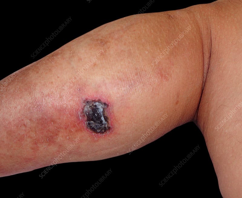
The condition typically begins as a small, painful pustule or nodule. This lesion breaks down rapidly and expands into a deep, irregular ulcer with a characteristic violaceous, meaning purple-blue, undermined border. The ulcer can grow alarmingly fast, sometimes expanding by several centimetres within days.
A Misunderstood Condition
For much of medical history, doctors misunderstood and misdiagnosed PG. Its name is a historical misnomer, coined before its true nature was understood. Today, clinicians recognise it as an immune-mediated condition, not an infectious one. This distinction is critical because treating it as an infection with antibiotics alone is ineffective and delays proper care.
PG is rare, affecting approximately three to ten people per million annually worldwide. It can affect people of any age, gender, or ethnicity, although it most commonly appears in adults between the ages of 40 and 60.
Types of Pyoderma Gangrenosum
Doctors recognise several clinical subtypes of PG. Each subtype has distinct features and may respond differently to treatment.
Ulcerative Pyoderma Gangrenosum
The ulcerative subtype is the most common form. It presents as a painful ulcer with a necrotic, meaning dead tissue, base and a raised, undermined, purple border. Ulcerative PG most often appears on the lower legs. This form carries the strongest association with IBD and other systemic diseases.
Pustular Pyoderma Gangrenosum
Pustular PG appears as discrete, painful pustules rather than large ulcers. These pustules are surrounded by redness and inflammation. This subtype associates strongly with IBD activity and tends to improve when the underlying bowel disease is treated effectively.
Bullous Pyoderma Gangrenosum
Bullous PG presents with superficial blisters, called bullae, rather than deep ulcers. It typically affects the face and upper limbs. This subtype associates with haematological conditions, particularly myeloid leukaemia and myelodysplastic syndrome.
Vegetative Pyoderma Gangrenosum
Vegetative PG is the mildest and least aggressive subtype. It presents as a superficial, warty plaque rather than a deep ulcer. Unlike other subtypes, it rarely associates with systemic disease and responds well to local treatment.
Peristomal Pyoderma Gangrenosum
Peristomal PG develops around stoma sites, the surgically created openings in the abdomen used in bowel surgery. This subtype is particularly relevant to people with IBD who have undergone colostomy or ileostomy procedures.
What Causes Pyoderma Gangrenosum?
The exact cause of PG remains incompletely understood. However, research points strongly toward immune system dysregulation as the central mechanism.
Immune System Dysregulation
In PG, neutrophils accumulate abnormally in the skin and release destructive enzymes and inflammatory chemicals. This creates a self-perpetuating cycle of tissue destruction. The trigger for this abnormal response remains unclear, but genetic and environmental factors both appear to contribute.
Recent research suggests that dysregulation of interleukin-1 (IL-1), interleukin-8 (IL-8), and tumour necrosis factor-alpha (TNF-α) plays a significant role. These are signalling molecules the immune system uses to coordinate inflammation. When their activity becomes dysregulated, excessive skin destruction follows.
The Role of Pathergy
A distinctive feature of PG is pathergy, an exaggerated skin response to minor trauma. In people with PG, a small wound, an insect bite, a needle puncture, or even surgical incision can trigger a new or worsening ulcer. This phenomenon makes wound care and any surgical procedures particularly challenging and risky.
Associated Systemic Conditions
PG rarely occurs in isolation. Around 50 to 70 percent of cases associate with an underlying systemic condition. IBD, including Crohn’s disease and ulcerative colitis, accounts for the most common association. Rheumatoid arthritis, haematological malignancies, and other autoimmune conditions also link with PG.
Identifying and treating the underlying condition often improves PG outcomes significantly.
The Connection Between Pyoderma Gangrenosum and IBD
The relationship between PG and IBD is one of the most clinically important aspects of this condition. PG represents one of the most common extraintestinal manifestations of IBD, meaning symptoms that occur outside the bowel.
How Common Is This Association?
Studies estimate that PG occurs in one to two percent of people with ulcerative colitis and a similar proportion of those with Crohn’s disease. While these percentages seem small, they represent thousands of people given the global prevalence of IBD. PG can appear before, during, or after IBD diagnosis, making the timeline unpredictable.
Does IBD Activity Drive PG?
The relationship between IBD disease activity and PG activity is complex. In some people, PG flares coincide with IBD flares. In others, PG behaves independently, even when IBD is in remission. This variability means doctors cannot rely on IBD control alone to manage PG in every patient.
Shared Inflammatory Pathways
Both IBD and PG involve dysregulated immune responses with overlapping inflammatory pathways. This explains why medications used to treat IBD, particularly biologics targeting TNF-α, also show effectiveness against PG. Treating the shared underlying immune dysfunction benefits both conditions simultaneously in many cases.
Gastroenterologists and dermatologists increasingly collaborate to manage patients who have both conditions, recognising that a coordinated approach produces better outcomes.
Signs and Symptoms of Pyoderma Gangrenosum
Recognising PG early is crucial because rapid progression can cause significant tissue damage and scarring.
Early Warning Signs
PG typically begins with a small, tender papule, pustule, or nodule on the skin. These early lesions are easy to dismiss as insect bites or minor skin infections. However, they rapidly deteriorate, breaking down into painful ulcers within days.
The pain associated with PG is often disproportionately severe compared to the lesion’s appearance. Intense, burning pain at the wound site is a hallmark feature that should prompt clinicians to consider PG as a diagnosis.
Established Ulcers
Once an ulcer forms, it typically displays several characteristic features. The base of the ulcer is necrotic, appearing grey or yellow with irregular, ragged edges. The border is raised, undermined, and distinctively purplish-blue. Surrounding skin is often red and inflamed.
Ulcers can range from one centimetre to more than 30 centimetres in diameter. Without treatment, they continue expanding relentlessly.
Systemic Symptoms
Some people with PG experience systemic symptoms alongside skin involvement. Fever, fatigue, and joint pain can accompany active PG. These symptoms reflect the systemic nature of the underlying inflammatory process.
When PG coexists with active IBD, bowel symptoms such as diarrhoea, abdominal pain, and rectal bleeding may also be prominent. This symptom overlap reinforces the importance of investigating for underlying systemic disease in every PG patient.
Diagnosing Pyoderma Gangrenosum
PG has no single definitive diagnostic test. Diagnosis relies on clinical assessment, exclusion of other conditions, and sometimes skin biopsy findings.
Clinical Diagnosis
Experienced dermatologists often diagnose PG primarily on clinical grounds. The characteristic ulcer appearance, rapid progression, severe pain, and pathergy response together create a recognisable clinical picture. A thorough history, including any known IBD or autoimmune conditions, strengthens the diagnosis.
Several diagnostic criteria frameworks exist. The Delphi consensus criteria, published in 2018, provide a structured approach to PG diagnosis, incorporating major and minor criteria based on clinical and histological features.
Skin Biopsy
Skin biopsy does not confirm PG definitively, but it serves an important purpose. Histological examination, meaning microscopic tissue analysis, typically shows dense neutrophilic infiltration in the dermis. More importantly, biopsy helps exclude other conditions that mimic PG, such as vasculitis, deep fungal infections, and cutaneous malignancies.
Doctors should ideally biopsy the active border of the ulcer rather than the necrotic base for the most informative results.
Investigations to Identify Underlying Disease
Every person newly diagnosed with PG requires investigation for underlying systemic conditions. Blood tests, colonoscopy, imaging, and bone marrow examination may all play a role depending on clinical suspicion. Identifying an underlying condition changes both prognosis and treatment strategy significantly.
Treatment of Pyoderma Gangrenosum
Treating PG requires a tailored, multi-pronged approach. Because no universal treatment exists, clinicians must adapt therapy to each individual’s disease severity, subtype, and associated conditions.
Topical and Local Therapies
Mild or localised PG may respond to potent topical corticosteroids or topical calcineurin inhibitors such as tacrolimus. Intralesional corticosteroid injections directly into the ulcer border can also help in early or limited disease.
Local wound care remains essential regardless of systemic treatment. Non-traumatic dressings, moisture balance, and infection prevention all support healing. Clinicians must take extreme care to avoid pathergy-inducing trauma during dressing changes.
Systemic Corticosteroids
Systemic corticosteroids, typically prednisolone, remain the most widely used first-line treatment for moderate to severe PG. High doses suppress the abnormal inflammatory response rapidly. Most people show improvement within weeks of starting treatment.
However, long-term corticosteroid use carries significant side effects, including weight gain, osteoporosis, diabetes, and infection susceptibility. Doctors aim to taper doses as quickly as safely possible.
Immunosuppressive Agents
When corticosteroids alone prove insufficient or cause unacceptable side effects, doctors add immunosuppressive medications. Ciclosporin, a powerful immune suppressant, shows good evidence for PG and can work rapidly. Dapsone, mycophenolate mofetil, and azathioprine are other options used in ongoing management.
These medications require careful monitoring for side effects including kidney dysfunction, liver toxicity, and increased infection risk.
Biologic Therapies
Biologic medications have transformed PG management, particularly for patients with coexisting IBD. TNF-alpha inhibitors, including infliximab and adalimumab, show strong evidence for PG treatment. Infliximab has demonstrated rapid ulcer healing in clinical trials.
Other biologics, including ustekinumab and secukinumab, are under investigation for PG. As understanding of PG’s inflammatory pathways deepens, targeted biologic therapy is likely to become increasingly central to management.
Wound Care and Surgical Considerations
Wound care in PG requires specialist expertise. Standard wound care principles must adapt to account for pathergy and the inflammatory nature of the ulcers.
Specialist Wound Management
Wound care nurses and dermatologists work together to select appropriate dressings that minimise trauma. Hydrogel dressings, foam dressings, and negative pressure wound therapy can support healing in suitable cases. Regular wound assessment monitors progress and detects secondary infection early.
Pain management during wound care is essential. Many people with PG require pre-medication with analgesics before each dressing change.
The Risks of Surgery
Surgery presents a significant challenge in PG because of pathergy. Surgical debridement, meaning removal of dead tissue, can paradoxically worsen ulcers by triggering new PG activity at the surgical site. For this reason, surgeons generally avoid aggressive debridement in active PG.
Skin grafting may benefit carefully selected patients with healing ulcers, but only after adequate systemic disease control. Timing and patient selection are critical.
Living With Pyoderma Gangrenosum
PG profoundly affects quality of life. Chronic pain, wound management demands, scarring, and the psychological burden of a visible, disfiguring condition all take a significant toll.
Psychological Impact
Many people with PG experience anxiety, depression, and social withdrawal. The unpredictable nature of the condition, combined with its visible impact, makes psychological support an important part of comprehensive care. Access to counselling, peer support groups, and mental health services improves overall outcomes.
Long-Term Outlook
PG can be a chronic, relapsing condition. Even after successful treatment, recurrence occurs in up to 30 percent of people. Long-term follow-up with a specialist dermatologist is essential. People with underlying IBD or other systemic diseases need coordinated multidisciplinary care to manage both the skin and systemic components of their illness.
With appropriate treatment, most ulcers eventually heal, though scarring is common. Early diagnosis and prompt treatment remain the most important factors in limiting long-term damage.
Frequently Asked Questions
What is Pyoderma Gangrenosum caused by?
Pyoderma Gangrenosum results from an abnormal immune response where neutrophils accumulate in the skin and cause tissue destruction. The exact trigger remains unclear. In most cases, PG associates with an underlying systemic condition such as inflammatory bowel disease, rheumatoid arthritis, or a blood disorder. Genetic predisposition and immune system dysregulation both contribute to its development.
Is Pyoderma Gangrenosum contagious?
No. Pyoderma Gangrenosum is not contagious. It is not caused by bacteria, viruses, or any infectious agent. It is an immune-mediated inflammatory condition. People cannot catch it from others, and it poses no infection risk to healthcare workers, family members, or caregivers.
How is Pyoderma Gangrenosum different from a regular ulcer?
Unlike ordinary skin ulcers caused by poor circulation or pressure, PG ulcers result from immune system attack on the skin. They expand rapidly, cause severe pain disproportionate to their size, and display a characteristic purple undermined border. They also show pathergy, worsening with minor trauma rather than healing as expected.
Can Pyoderma Gangrenosum be cured?
There is no guaranteed cure for PG. However, many people achieve complete ulcer healing with appropriate treatment. Recurrence remains possible, particularly if underlying systemic disease is not well controlled. Long-term management focusing on immune suppression and treatment of associated conditions offers the best chance of sustained remission.
Does treating IBD help Pyoderma Gangrenosum?
In many cases, yes. Because PG and IBD share overlapping inflammatory pathways, medications used to treat IBD, particularly biologic therapies, can also improve PG. However, the relationship is not consistent. Some people experience PG flares even when their IBD is in remission, requiring PG-specific treatment independently of bowel disease management.
How long does Pyoderma Gangrenosum take to heal?
Healing times vary considerably. With appropriate treatment, some ulcers begin improving within weeks. Others take months to heal fully. Larger ulcers, delayed diagnosis, and inadequate treatment all prolong healing. Even after healing, significant scarring often remains. Regular specialist follow-up is essential throughout the healing process.
Disclaimer:
This article is for informational purposes only and does not constitute medical advice. Always consult a qualified healthcare professional for diagnosis, treatment, or medical guidance related to any health condition.
References:
Observer Voice is the one stop site for National, International news, Sports, Editor’s Choice, Art/culture contents, Quotes and much more. We also cover historical contents. Historical contents includes World History, Indian History, and what happened today. The website also covers Entertainment across the India and World.