

Microscopic Colitis: The Bowel Inflammation You Can’t See with a Camera

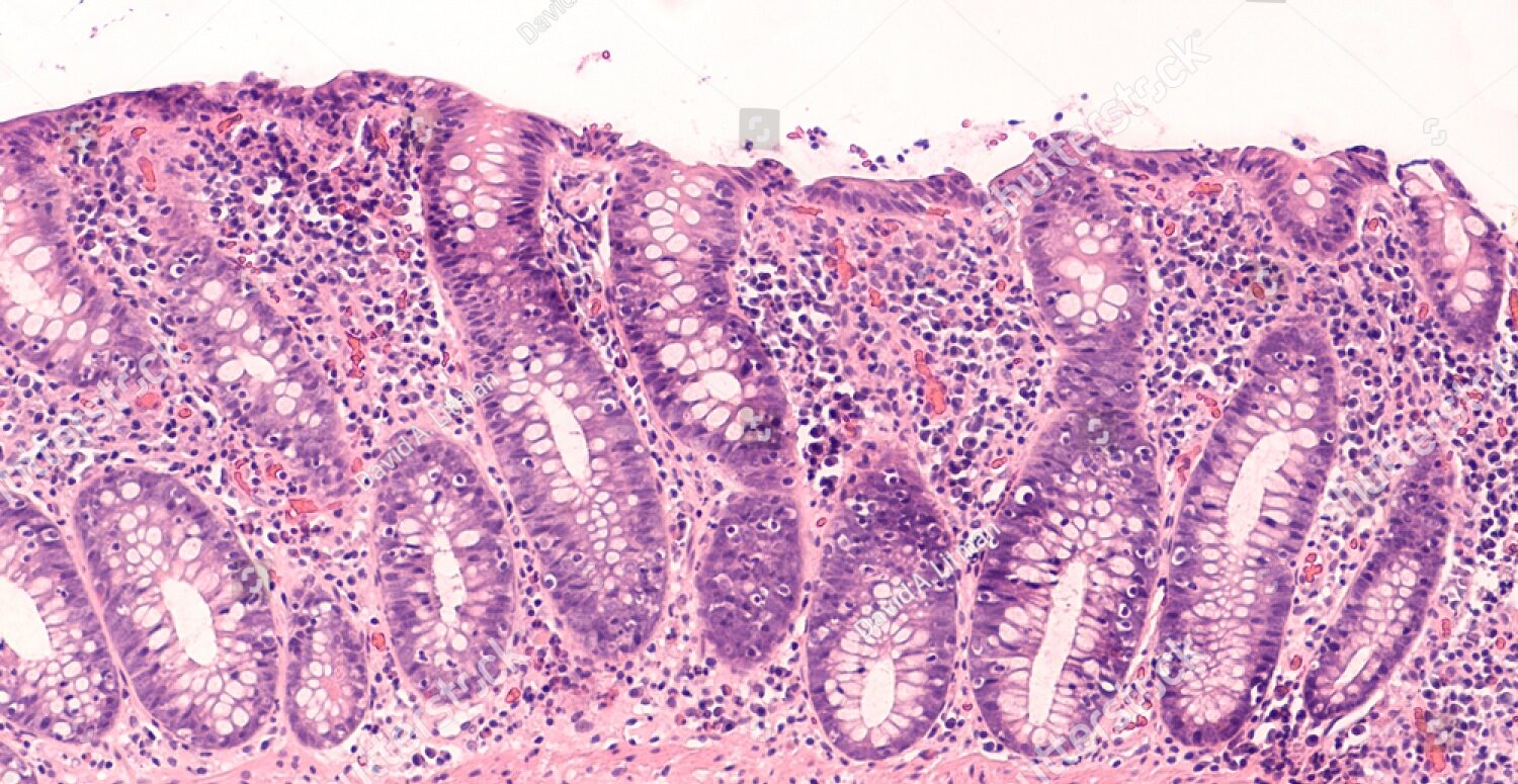
If you have been running to the bathroom ten times a day with watery diarrhea, but your colonoscopy came back completely normal, you are not imagining things. There is a real condition that hides in plain sight — one that a camera simply cannot detect. Understanding what is microscopic colitis may be the key to finally getting the right diagnosis and the relief that has been out of reach.
Microscopic colitis is an inflammatory bowel disease where doctors can only see the inflammation by looking at colon tissue under a microscope. The colon itself appears perfectly healthy during a standard endoscopy. Research suggests that in the United States alone, approximately 700,000 people have microscopic colitis, and the disease is three to nine times more common in women than in men. Despite those numbers, it is routinely misidentified as irritable bowel syndrome or functional diarrhea, leaving patients without effective treatment for months or years. NIDDKNIDDK
What Is Microscopic Colitis
Increasingly considered an inflammatory bowel disease, microscopic colitis is a cause of chronic watery, non-bloody diarrhea diagnosed predominantly in older patients. It has three major histological subtypes: collagenous colitis, lymphocytic colitis, and incomplete microscopic colitis. The word “microscopic” does not mean the disease is minor. It simply describes how it is detected. The only way a doctor can diagnose it is to take a sample of tissue and check it under a microscope. NCBIWebMD
The colon typically appears normal endoscopically, and the diagnosis requires histologic evaluation. This is what makes the condition so easy to miss. A patient can undergo a full colonoscopy, receive a normal result, and still be living with significant bowel disease. Without a doctor who specifically requests tissue biopsies, the diagnosis is never made. Mayo Clinic Proceedings
The Two Main Types of Microscopic Colitis
In lymphocytic colitis, the colon lining contains more white blood cells than normal, while the layer of collagen under the colon lining is normal or only slightly thicker than normal. In collagenous colitis, the layer of collagen under the colon lining is thicker than normal, and the colon lining may also contain more white blood cells than normal. Both types produce the same symptoms and are treated in the same way, so the clinical distinction matters more to the pathologist examining the tissue than to the patient experiencing the symptoms. NIDDK
For collagenous colitis, the female-to-male ratio can range from 4.4 to 7.9 to one, while for lymphocytic colitis it ranges from 1.8 to 5.0 to one. A third form, incomplete microscopic colitis, describes cases where tissue changes are present but do not fully meet the diagnostic thresholds for either major subtype. It produces similar symptoms and is managed along the same lines. Biology Insights
How Common Is Microscopic Colitis
A meta-analysis indicated a worldwide incidence of approximately 4.9 cases per 100,000 person-years for collagenous colitis and 5.0 cases per 100,000 person-years for lymphocytic colitis. Overall prevalence has been reported at around 103 per 100,000 persons. Those numbers have been climbing steadily. A Danish nationwide study observed a tenfold increase in incidence from 2.3 cases per 100,000 person-years in 2001 to 24.3 cases per 100,000 person-years by 2016, and this rise has led to microscopic colitis surpassing the incidence rates of Crohn’s disease and ulcerative colitis in some countries. Biology InsightsBiology Insights
The overall incidence rate is estimated at 11.4 cases per 100,000 person-years, with a higher incidence in adults older than 60 years and in women. The predominance among older women has not been fully explained but likely reflects a combination of age-related immune dysregulation, a greater burden of autoimmune diseases, patterns of medication exposure, and potential hormonal influences. Despite its growing prevalence, public awareness of the condition remains strikingly low. Clinical Advisor
Causes and Risk Factors
The exact cause of microscopic colitis is not fully understood, and researchers believe it is multifactorial — meaning several things likely work together to trigger it, particularly in people who are already genetically predisposed. Experts think abnormal reactions of the immune system may play a role, leading to inflammation in the colon. People who have certain immune disorders such as celiac disease, rheumatoid arthritis, or type 1 diabetes are more likely to develop microscopic colitis, and research suggests certain genes also increase the chance a person will develop the condition. NIDDK
Proton pump inhibitors, nonsteroidal anti-inflammatory drugs, selective serotonin reuptake inhibitors, and statins have all been associated with microscopic colitis in observational studies. PPIs modify gastric and intestinal pH, which can disrupt the bowel microbiome. Importantly, not everyone who takes these medications will develop the condition. They appear to act as triggers in people who are already susceptible. Multiple prospective cohort and cross-sectional studies have shown a strong association between cigarette smoking and microscopic colitis, with an odds ratio of 2.12, and a significantly higher risk among current smokers compared with never-smokers. Tricyclic antidepressants, commonly prescribed for functional gastrointestinal disorders, have also been identified as an emerging risk factor in recent cohort research. Patients should never stop a prescribed medication without first consulting their doctor. Clinical Advisornih
The incidence was higher in women than in men and increased with older age, demonstrating a peak between 60 and 80 years of age, in particular in postmenopausal women on hormone replacement therapy and also in women using an oral contraceptive. This hormonal dimension adds another layer of complexity to a condition that already has multiple contributing pathways. frontiersin
Symptoms of Microscopic Colitis
The most common symptom of microscopic colitis is chronic, non-bloody, watery diarrhea. Other symptoms may include fecal urgency, nocturnal stools, abdominal pain, arthralgia, weight loss, and fecal incontinence. The severity of these symptoms is variable, and sometimes they can be misinterpreted as diarrhea-predominant irritable bowel syndrome, leading to inadequate treatment. NCBI
The non-bloody nature of the diarrhea is a critical distinguishing detail. Unlike ulcerative colitis, which typically produces visible blood in the stool, microscopic colitis does not. Nocturnal diarrhea — being woken from sleep by the urgent need to use the bathroom — is a particularly telling sign that something more than IBS is at play, since true IBS rarely disturbs sleep. Patients with active disease present with more than three stools daily or more than one watery stool every day. Dehydration, fatigue, and unintended weight loss can develop over time when the condition is left untreated. The unpredictability of flares creates real anxiety and can severely limit daily life, social activities, and professional functioning. NCBI
How Microscopic Colitis Is Diagnosed
No laboratory tests can establish this diagnosis. These patients must undergo colonoscopy with biopsy. The pathologist usually classifies patients as having lymphocytic colitis, with increased numbers of lymphocytes in the mucosa, or collagenous colitis, with an increase in collagen deposition in the submucosa. nih
Since the typical histological changes in microscopic colitis can be patchy, it is crucial to take biopsies from all colon regions to allow the pathologist to make the diagnosis. It is recommended to harvest at least two biopsies from the ascending colon, the transverse colon, the descending colon, the sigmoid colon, and the rectum, in order to achieve a diagnostic sensitivity greater than 95%. This is precisely why the condition is so often missed. If a colonoscopist does not take systematic biopsies across the full length of the colon, disease in an unsampled segment will simply go undetected. Patients who receive a normal colonoscopy result but continue to experience severe diarrhea should ask their gastroenterologist specifically whether multiple biopsies were collected from different parts of the colon. Blood tests and stool cultures are used to rule out other causes, including infection, thyroid disorders, and celiac disease, but the biopsy remains the definitive diagnostic tool. nih
Treatment and Management
Medicines most often treat microscopic colitis effectively. Doctors prescribe medicines to improve symptoms and achieve remission — a time when symptoms improve or disappear. The first step is identifying and removing any medication that may be acting as a trigger. If a proton pump inhibitor, NSAID, or SSRI is suspected, the prescribing doctor may adjust or substitute it. Stopping smoking is an equally important early intervention. NIDDK
Budesonide is the recommended initial treatment. It is a poorly absorbed oral corticosteroid medication that works locally within the colon lining, reducing inflammation with fewer systemic side effects than conventional steroids. Most patients begin with an eight-week induction course. Because relapse after stopping is common, maintenance therapy is frequently required, and the decision to extend treatment is made on an individual basis by the treating gastroenterologist. nih
Antidiarrheal medications such as loperamide or bismuth subsalicylate may be sufficient in patients with mild symptoms. Bile acid sequestrants such as cholestyramine are useful when abnormal bile acid absorption is contributing to loose stools. For patients who do not respond adequately to budesonide, options include aminosalicylates, immunosuppressants, and biologics. Recent studies have demonstrated promising efficacy of vedolizumab and tumor necrosis factor inhibitors in the treatment of steroid-refractory microscopic colitis. Surgery is extremely rare and reserved only for the most severe treatment-resistant cases. On the dietary side, reducing caffeine, alcohol, artificial sweeteners, and high-fat foods during active flares helps manage symptoms, and a gluten-free diet may provide added benefit in patients who also have celiac disease. CghjournalCghjournal
Prevention and Long-Term Outlook
There is no guaranteed way to prevent microscopic colitis, but practical steps can meaningfully reduce personal risk. Using NSAIDs only when necessary, discussing long-term PPI alternatives with a doctor, and quitting smoking are the most evidence-supported interventions available. For people already diagnosed, identifying and eliminating individual triggers — whether dietary, pharmaceutical, or lifestyle-related — remains the most effective strategy for reducing the frequency and severity of flares.
Whereas recent studies suggest that the incidence of microscopic colitis has plateaued, given the aging population, the prevalence will likely increase. Careful evaluation of exacerbating factors is essential in management, and given the high risk of relapse, maintenance therapy is often required. On the question of cancer risk, current evidence is reassuring. Microscopic colitis does not make a person more likely to get colorectal cancer, which distinguishes it meaningfully from ulcerative colitis and Crohn’s disease. Some population studies have noted a modest association with lymphoma and lung cancer, but these findings have not yet changed standard clinical management. Mayo Clinic ProceedingsWebMD
Latest Research and Updates
The research landscape has advanced considerably in recent years. A major 2024 update published in Clinical Gastroenterology and Hepatology reviewed new randomised trial data and offered updated guidance on biologics for patients whose disease does not respond to steroids. Early results for vedolizumab, a gut-selective biologic already approved for other forms of inflammatory bowel disease, are particularly promising for steroid-refractory microscopic colitis.
Because biomarkers are absent in microscopic colitis, the diagnosis relies on colonoscopy with histological assessment of biopsy specimens from all parts of the colon. Scientists are actively working to identify non-invasive diagnostic tools — including fecal calprotectin and other stool markers — that could eventually reduce the need for colonoscopy in every suspected case, though none has yet reached the accuracy required to replace histology in routine practice. Bone health has also emerged as an important management consideration. Long-term budesonide use in older women already at elevated risk for osteoporosis requires periodic monitoring of bone density, and some studies suggest microscopic colitis may itself independently contribute to reduced bone density. For further reading on digestive health and inflammatory bowel conditions, visit ObserverVoice.com. nih
Key Takeaways
Microscopic colitis is a genuine, diagnosable inflammatory bowel disease that causes persistent watery diarrhea while producing no visible abnormalities on a standard colonoscopy. It is more common than most people realise, disproportionately affects older women, and is strongly linked to several widely prescribed medications including PPIs, NSAIDs, and SSRIs. Diagnosis depends entirely on tissue biopsies collected systematically from multiple segments of the colon and examined under a microscope. Budesonide is the proven first-line treatment, and most patients can achieve meaningful remission. The condition does not increase colorectal cancer risk. With the correct diagnosis and an individualised treatment plan, life with microscopic colitis is manageable.
Frequently Asked Questions
Can microscopic colitis go away on its own?
In some cases, symptoms resolve spontaneously, particularly after a triggering medication is identified and discontinued. However, the condition is often relapsing in nature, and many patients require medical treatment to achieve and maintain remission. A gastroenterologist should continue monitoring progress even when symptoms appear to have settled, as silent disease activity can persist.
Is microscopic colitis the same as IBS?
No. While the two conditions share surface-level symptoms such as chronic diarrhea and cramping, they are fundamentally different diseases. IBS produces no abnormal findings on biopsy, whereas microscopic colitis causes identifiable structural changes in colon tissue that are only visible under a microscope. Patients with microscopic colitis are frequently misdiagnosed with IBS before appropriate biopsies are carried out.
Does microscopic colitis increase cancer risk?
Current evidence does not show an increased risk of colorectal cancer in people with microscopic colitis, which distinguishes it favourably from ulcerative colitis and Crohn’s disease. Some population-based cohort studies have noted a modest association with lymphoma and lung cancer, but these findings have not yet translated into any change in standard clinical surveillance guidelines.
What foods should I avoid during a flare?
Caffeine, alcohol, dairy products, artificial sweeteners, and high-fat or high-fibre foods are among the most commonly reported dietary triggers. Individual tolerance varies considerably, and no single diet has been proven effective for all patients. Keeping a detailed food and symptom diary helps identify personal triggers. A gluten-free diet may offer particular benefit in patients who also have underlying celiac disease.
How long will I need to take budesonide?
Most treatment courses begin with an eight-week induction course of budesonide. Because relapse after stopping is common, gastroenterologists often recommend a tapered schedule or longer maintenance therapy. The appropriate duration depends on how frequently symptoms return, how well the medication is tolerated, and the individual patient’s clinical picture. Long-term use in older patients requires periodic bone density monitoring to guard against steroid-related bone loss.
References
Disclaimer: This article is based on publicly available information from the National Institutes of Health, the National Institute of Diabetes and Digestive and Kidney Diseases, and peer-reviewed medical literature. It is intended for informational and educational purposes only and does not constitute medical advice. ObserverVoice.com is a news and information platform and not a healthcare provider. Always consult a qualified medical professional for personal diagnosis and treatment decisions.
Observer Voice is the one stop site for National, International news, Sports, Editor’s Choice, Art/culture contents, Quotes and much more. We also cover historical contents. Historical contents includes World History, Indian History, and what happened today. The website also covers Entertainment across the India and World.

