

Osteogenesis Imperfecta (Brittle Bone Disease): Living With Bones That Break Easily

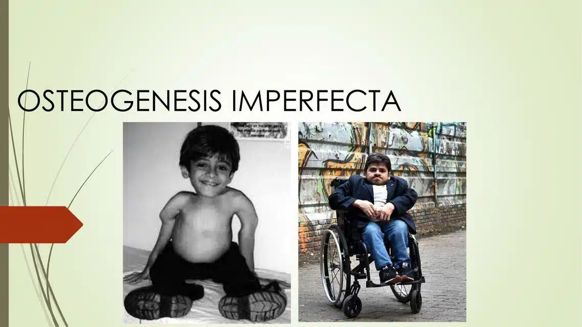
Imagine a child who fractures bones repeatedly. A fall from a height causes multiple fractures. A minor bump causes a break. Growth hurts. Bones break during normal childhood activities. The diagnosis is osteogenesis imperfecta—brittle bone disease. A genetic disorder causing defective collagen production, resulting in extremely weak bones. The family fears a future of constant fractures, disability, and dependence. Yet modern bisphosphonate treatment, physical therapy, genetic counseling, and supportive care have transformed osteogenesis imperfecta from a severely limiting condition to one compatible with school attendance, sports participation, and meaningful adult lives. People with osteogenesis imperfecta become athletes, artists, professionals, and leaders. Fracture prevention through medication and therapy reduces fracture frequency. Surgical intervention stabilizes bones. Psychological support enables coping. Understanding osteogenesis imperfecta enables appropriate care and realistic expectations. Osteogenesis imperfecta is a genetic disorder caused by mutations in genes encoding type I collagen, the primary structural protein in bone. Defective collagen production results in extremely fragile bones prone to fractures from minor trauma. The condition is autosomal dominant inheritance. Approximately 50 percent of cases are inherited. Approximately 50 percent are de novo mutations. Osteogenesis imperfecta affects approximately 1 in 10,000 to 1 in 20,000 people. Approximately 20,000 to 50,000 Americans have OI. The condition has multiple types varying in severity. Type I—mild. Type II—perinatal lethal. Type III—severe progressive. Type IV—moderate to severe. What makes osteogenesis imperfecta important is understanding the range of severity. Type II is fatal. Type I is mild with near-normal lifespan. Types III and IV require intensive management but enable meaningful living. Modern bisphosphonate treatment increases bone density, reduces fracture frequency, and improves outcomes dramatically. Early intervention prevents complications. Understanding osteogenesis imperfecta enables appropriate medical management and realistic life planning. In this comprehensive article, we will explore what osteogenesis imperfecta is, understand the collagen mutations, recognize the types and severity spectrum, explore fracture complications and management, and discover how modern treatment enables full, active participation in life.
Understanding Type I Collagen Function and OI Pathophysiology
Before we explore osteogenesis imperfecta, we need to understand type I collagen and how mutations cause bone fragility. Type I collagen. Primary structural protein. Bone. Skin. Tendons. Ligaments. Sclera. Teeth. Comprises approximately 90 percent of bone organic matrix. Provides tensile strength. Bone resistance to pulling forces. Hydroxyapatite minerals. Provide compressive strength. Bone resistance to compression. Balance. Strong bones. Collagen architecture intact. Mineral density appropriate. Together. Optimal bone strength. Type I collagen structure. Triple helix. Three polypeptide chains. Collagen alpha-1(I). Collagen alpha-2(I). Wound around each other. Stabilized by hydrogen bonds. Cross-links. Between molecules. Create network. Fiber bundles. Organized structure. Strength. Flexibility. Type I collagen genes. COL1A1 gene. Encodes pro-alpha1(I) chain. COL1A2 gene. Encodes pro-alpha2(I) chain. Located on chromosomes 17 and 7. Different locations. COL1A1 mutations. Approximately 90 percent of OI. COL1A2 mutations. Approximately 10 percent of OI. Different mutation types. Different severity. Collagen synthesis. Procollagen. Pro-alpha chains synthesized. Hydroxylation. Proline to hydroxyproline. Lysine to hydroxylysine. Requires vitamin C. Cofactor. Triple helix formation. Three chains. Hydrogen bonded. Stabilized. Post-translational modifications. Cross-linking. Lysyl oxidase. Converts lysine to allysine. Cross-links form. Between molecules. Secretion. Collagen secreted. Extracellular matrix. Assembly. Collagen fibrils form. Organization. Fiber bundles. Bone matrix formation. In OI. Collagen mutations. Defective synthesis. Defective structure. Defective assembly. Defective secretion. Different mutations. Different effects. Haploinsufficiency. One normal copy. One mutated copy. Insufficient collagen. Reduced quantity. Normal collagen. But not enough. Structural mutation. Both normal and mutant collagen. Mutant distorts helix. Weakens overall structure. Dominant negative. Small amount mutant. Affects much more. Causes severe defects. Two-hit hypothesis. Not applicable. Heterozygous. One mutated copy. Sufficient for disease. Homozygous. Two mutated copies. Usually embryonic lethal. Type II. Lethal in utero. Or perinatally. Dominantly inherited. One mutated copy. Sufficient for severe disease. OI types. Classification system. Based on severity. Clinical features. Genetic basis. Helps predict prognosis. Guides management. Type I. Mild OI. Autosomal dominant. COL1A1 haploinsufficiency. Reduced collagen quantity. Normal structure. Fragile bones. Frequent fractures. But not severe. Joints. Usually normal mobility. Sometimes hypermobility. Sclera. Blue-tinged sometimes. Teeth. Normal usually. Dentinogenesis imperfecta. Sometimes. Hearing. Normal usually. Hearing loss. Sometimes. Progressive. Lifespan. Near-normal. Approximately normal. Type II. Perinatal lethal OI. Autosomal dominant. COL1A1 or COL1A2 mutations. Structural defects. Dominant negative. Severe collagen defects. Extremely fragile bones. Multiple in utero fractures. Severe skeletal deformities. Blue sclera. Characteristic. Severe hypomineralization. Lethal in utero. Or perinatally. Usually first few days. Severe respiratory complications. Cardiac abnormalities. Renal abnormalities. Incompatible with life. Type III. Severe progressive OI. Autosomal dominant. COL1A2 mutations. Predominant. Severe collagen defects. Extremely fragile bones. Multiple fractures. Progressive deformities. Sclera. Bluish. Teeth. Dentinogenesis imperfecta. Hearing loss. Progressive. Joint hypermobility. Severe. Progressive kyphoscoliosis. Severe spinal deformity. Growth. Severe growth restriction. Wheelchair dependence. Often. Many fractures. Frequency. Lifespan. Shortened. Complications. Mortality. Early adulthood. Sometimes. Type IV. Moderate to severe OI. Autosomal dominant. COL1A1 or COL1A2 mutations. Intermediate phenotype. Between type I and III. Fragile bones. Multiple fractures. Progressive deformities. Sclera. Normal white. Distinguishes from types I-III. Teeth. Normal usually. Hearing loss. Possible. Joint hypermobility. Variable. Kyphoscoliosis. Progressive. Growth. Near-normal to moderately restricted. Ambulatory. Wheelchair. Variable. Lifespan. Near-normal. Approximately normal. Other types. Type V. Autosomal dominant. IFITM5 gene. Interferon-induced transmembrane. Collagen quantity. Normal. Collagen quality. Normal. Mechanism. Unknown. Bone defects present. Dense metaphyseal bands. Radiographic finding. Calcification. Interosseous membrane. Forearm. Restricted pronation. Hypermobility. Limited. Type VI. Autosomal recessive. SERPINF1 gene. Serine protease inhibitor clade F. Collagen defects. Bone defects. Moderate. Type VII. Autosomal recessive. CRTAP gene. Cartilage-associated protein. Collagen defects. Severe phenotype. Type VIII and beyond. Rare. Various genes. Autosomal recessive. The pathophysiology explains the bone fragility and complications in OI.
What is Osteogenesis Imperfecta?
Osteogenesis imperfecta is a genetic disorder caused by type I collagen mutations resulting in extremely fragile bones prone to fractures, bone deformities, and various systemic complications. Type I OI. Mild form. Autosomal dominant. COL1A1 haploinsufficiency. Most common type. Approximately 85 percent of OI. Clinical features. Blue sclera. Characteristic. Bone fragility. Fractures common. But not severe. Variable. Some have few fractures. Some have many. Dental problems. Dentinogenesis imperfecta. Approximately 50 percent. Enamel defects. Discoloration. Wearing. Progressive decay. Hearing loss. Progressive. Approximately 25 to 50 percent. Adolescent or adult. Conductive or sensorineural. Joint hypermobility. Usually mild. Skin fragility. Easy bruising. Variable. Healing. Normal usually. Lifespan. Near-normal. Intelligence. Normal. No cognitive impairment. Type II OI. Perinatal lethal. Most severe. Autosomal dominant. COL1A2 mutations. Dominant negative. Severe collagen defects. Clinical features. Severe skeletal dysplasia. Deformities. Multiple in utero fractures. Hypomineralization. Extreme. Blue sclera. Characteristic. Pathologic fractures. In utero. During delivery. Perinatally. Respiratory complications. Chest wall abnormalities. Restrictive lung disease. Respiratory failure. Common. Cardiac. Heart defects. Cardiac complications. Renal. Renal abnormalities. Renal failure. Prognosis. Lethal. In utero or perinatally. Usually first few hours or days. Incompatible with life. Type III OI. Severe progressive. Autosomal dominant. COL1A2 mutations. Structural defects. Clinical features. Severe bone fragility. Multiple fractures. Progressive deformities. Blue sclera. Characteristic. Dentinogenesis imperfecta. Common. Hearing loss. Progressive. High frequency. Approximately 50 to 75 percent. Joint hypermobility. Severe. Kyphoscoliosis. Progressive spinal deformity. Severe. Growth restriction. Short stature. Very short. Progressive deformity. Wheelchair dependence. Usually. Many fractures. Hundreds. Lifetime. Progressive disability. Prognosis. Shortened lifespan. Early adulthood. Complications. Mortality. Type IV OI. Moderate to severe. Autosomal dominant. COL1A1 or COL1A2. Intermediate phenotype. Clinical features. Fragile bones. Multiple fractures. Progressive deformities. White sclera. Usually. Normal or near-normal. Distinguishes from types I-III. Dentinogenesis imperfecta. Variable. Hearing loss. Possible. Progressive. Joint hypermobility. Variable. Kyphoscoliosis. Progressive. Short stature. Moderate to severe. Variable. Ambulatory or wheelchair. Variable. Prognosis. Near-normal lifespan. Approximately normal. Complications. Progressive deformity. Mobility restrictions. Joint problems. Arthritis. Hearing loss. Dental problems. Management. Similar to type III. But usually less severe. Fracture characteristics. Location. Any bone. Long bones—femur, tibia, humerus. Most common. Fracture frequency. Type I. Few fractures. Some never fracture. Others have multiple. Type III. Many fractures. Type IV. Many fractures. Healing. Normal usually. Callus formation. Sufficient usually. Non-union. Rare. Union. Usually rapid. Time. Often faster than population. Overhealing. Exuberant callus. Sometimes. Other complications. Basilar invagination. Skull base. Invagination into brainstem. Neurologic complications. Serious. Requires monitoring. Hydrocephalus. Ventricular enlargement. Increased intracranial pressure. Shunt placement. If symptomatic. Cardiomyopathy. Heart muscle weakness. Rare. Serious. Aortic regurgitation. Aortic valve. Valve insufficiency. Rare. Serious. Renal. Nephrolithiasis. Kidney stones. Increased risk. Genitourinary abnormalities. Variable. The systemic nature requires comprehensive surveillance.
Recognizing Osteogenesis Imperfecta: Fracture Patterns and Clinical Features
Osteogenesis imperfecta has distinctive features recognizable from infancy through adulthood. Infancy and early childhood (0 to 5 years). Birth presentation. Type I. Often undiagnosed at birth. May present later. With fractures. Type II. Obvious at birth. Severe skeletal dysplasia. Blue sclera. Severe deformities. Multiple fractures. Respiratory distress. Type III. At birth or early infancy. Blue or dark sclera. Obvious features. Type IV. May be undiagnosed initially. Fractures with trauma. Fractures in infancy. Unexplained. Concerning. Abuse evaluation. Important. Rule out non-accidental trauma. Genetic testing. Confirms OI diagnosis. Relieves suspicion. Fractures. First fractures. Often early. Infancy or early childhood. Clavicle fractures. Common first fracture. Birth trauma. Difficult delivery. Or spontaneous. Other bones. Femur. Tibia. Humerus. Fractures from minor trauma. Falls from height. Minor bumps. Handling. Even gentle. Can cause fractures. Healing. Rapid usually. Callus formation. Strong usually. Full healing. Weeks. Type I. Usually 6 to 8 weeks. Type III. Usually 6 to 8 weeks. Sometimes faster. Multiple fractures. Accumulate. Over months and years. Deformity from repeated fractures. Develops. Progressive. Bowing. Limbs. Anterior bowing. Tibia particularly. Posterior bowing. Femur. Sometimes. Kyphoscoliosis. Spinal deformity. Progressive. Type III particularly. Severe. Type I. Usually mild. Type IV. Moderate. Growth restriction. Type III particularly. Very short stature. Growth curve. Below normal. Progressive restriction. Type I. Usually near-normal growth. Type IV. Moderate restriction. Type II. Lethal. Non-viable. Sclera. Blue tinted. Types I, III, IV with collagen defects. Structural mutations. Type I. Sometimes. Not always. Type II. Always. Type III. Always. Type IV. Sometimes. White sclera. Does not rule out OI. Type IV. Sclera white. But OI present. Teeth. Dentinogenesis imperfecta. Discolored. Brown or yellow. Enamel. Defective. Wearing away. Progressive decay. Sensitive teeth. Early intervention. Dental care. Fluoride. Protect remaining structure. Type I. Approximately 50 percent. Type III. Common. Type IV. Variable. Type II. Usually. Hearing. Loss progressive. Types I, III, IV. Approximately 25 to 50 percent develop. Conductive—ossicular involvement. Stapes footplate. Fixation. Sensorineural—cochlear. Inner ear involvement. Speech development. Affected. Hearing aids. May help. Cochlear implant. Possible. Delayed speech. From hearing loss. Support. Speech therapy. Education. Important. Joints. Hypermobility. Joint laxity. Excessive motion. Subluxation. Joint dislocation. Risk. Musculoskeletal. Muscle strength. Weak sometimes. Type III particularly. Physical therapy. Strengthening. Helps. Mobilization. Functional movement. Improves. Bruising. Easy. Blue skin. Ecchymoses. Minor trauma. Disproportionate bruising. Fragility. Skin. Collagen affected. Healing. Normal usually. But bruising. Prominent. Appearance. Cosmetic concern. School age (5 to 12 years). Fractures continue. Type III. Frequent. Weekly or monthly. Type I. Variable. Some never fracture. Others have several. Type IV. Regular. Prevention. Medication. Bisphosphonates. Reduce fracture frequency. Approximately 30 to 50 percent reduction. Life-changing. Exercise. Physical therapy. Strengthening. Joint protection. Mobility. Maintained. Functional. Activity participation. Important. School. Integration. Regular school. Classroom. Usually. Accessibility. Important. Building. Ramps. Elevators. Accessible bathrooms. Classroom. Adapted. Desk height. Physical education. Modified. Swimming. Excellent. Weight-bearing. Controlled. Impact sports. Avoided. Academics. Normal. Intelligence. Average. Learning disabilities. Not from OI. Academic achievement. Possible. Full potential. Peer relationships. Fractures. School absences. Social impact. Support. Important. Body image. Different. Mobility. Crutches. Wheelchair. Variables. Acceptance. Peer support. Important. Psychological. Anxiety. About fractures. Normal. Counseling. Coping strategies. Helpful. Adolescence (12 to 18 years). Fractures continue. Type III. Frequent. Type I. Variable. Type IV. Regular. Growth. Puberty. Growth spurt. Fracture risk. Increases. Bone formation. Active. Rapid. Bisphosphonate. Important. Reduces fracture. Growth completion. Skeletal maturity. Variable age. Females usually earlier. Males usually later. Deformity. Progressive. Limb bowing. Worsening. Kyphoscoliosis. Progression. Functional impact. Mobility. Wheelchair. Increasing need. Type III particularly. Independence. Developing. Important. Autonomy. Physical assistance. Variable. Peer relationships. Dating. Social. Acceptance important. Sexuality. Sexual function. Usually normal. Conception. Possible. Genetic risk. Offspring risk. 50 percent if carrier. Genetic counseling. Important. Education. College. Possible. Accommodations. Accessibility. Support services. Important. Career planning. Exploration. Abilities. Interests. Realistic assessment. Occupational limitations. Variable. Work disability. Type III. Significant. Type I. Minimal. Type IV. Moderate. Adulthood (18+ years). Fractures continue. Risk. Lifetime. Type I. Lower. Type III. Higher. Type IV. Moderate. Bone density. Declines. Age. Osteoporosis. Risk. Especially postmenopausal women. Management. Calcium. Vitamin D. Weight-bearing exercise. Bisphosphonates. Continued. Into adulthood. Benefits. Debate. But widely used. Mobility. Type I. Usually independent. Type III. Wheelchair dependent. Type IV. Variable. Independence. Employment. Many employed. Meaningful work. Type I. Usually. Type III. Variable. Type IV. Usually. Relationships. Marriage. Family. Possible. Parenting. Ability. Depends on mobility. Support. Possible. Pregnancy. Increased fracture risk. During and after. Genetic counseling. Important. Offspring risk. 50 percent. Joint problems. Arthritis. Progressive degeneration. Pain. Management. NSAIDs. Physical therapy. Joint replacement. Possible. Hearing loss. Progressive. Hearing aids. Cochlear implant. Technology. Helps. Dental problems. Progressive. Tooth loss. Dentures. Implants. Options. Psychological. Adjustment. Chronic condition. Chronic pain. Frequency. Chronic disability. Psychological impact. Depression. Anxiety. Support. Counseling. Medication. Important. Quality of life. High priority. Despite physical challenges. Many achieve. Fulfilling lives. Meaningful activities. Relationships. Work. The diversity of presentations requires individualized management.
Diagnosis: Recognizing Osteogenesis Imperfecta
Diagnosing osteogenesis imperfecta requires clinical recognition, imaging findings, and genetic testing. Clinical features. Blue sclera. Distinctive. Types I, III, IV with mutations causing collagen defects. Type IV—white sclera. Still possible. Repeated fractures. Minor trauma. Excessive fracture frequency. For age. Concerning. Suggests OI. Family history. Family member with OI. Known. Genetic predisposition. Genetic testing. OI suspected. Dental problems. Dentinogenesis imperfecta. Discoloration. Enamel defects. Associated with OI. Hearing loss. Progressive. Adolescent. Suggests OI. Hypermobility. Joint laxity. Associated. OI. Clinical assessment. Physician evaluation. History. Fracture frequency. Age at first fracture. Circumstances. Trauma. Minimal. Or none. Radiographic findings. X-rays. Fractures. Healing. Callus. Ossification. Assessment. Bone density. Often low. Hypomineralization. X-rays appear. Osteopenic. Transparent. Metaphyseal features. Metaphyseal bands. Dense or lucent. Vertebral features. Biconcave vertebrae. Vertebral compression. Spine curvature. Kyphosis. Lordosis. Scoliosis. Skeletal survey. Comprehensive. Multiple bones. Assess severity. Assess deformities. DXA scan. Bone density measurement. T-score. Z-score. Assess. Percentage of expected. Baseline. Periodic. Monitor treatment. Genetic testing. DNA sequencing. COL1A1 gene. COL1A2 gene. Other genes. IFITM5. CRTAP. Others. Identifies mutation. Confirms diagnosis. Pathogenicity classification. Pathogenic mutations. Clearly cause OI. Benign variants. No disease. Variants of uncertain significance (VUS). Unknown. Requires research. Biochemical testing. Collagen synthesis. Sometimes tested. Fibroblast culture. Collagen analysis. Electrophoresis. Identifies mutations. Not routine. Research primarily. Collagen analysis. Skin biopsy. Culture. Type I collagen analysis. Mutation detection. Some centers. Molecular genetics. Direct testing. Now more common. More available. Diagnosis criteria. Clinical features. Plus genetic testing. Confirms OI. Genetic counseling. Before and after testing. Important. Discussion of inheritance. Offspring risk. Medical management. Genetic testing. Family members. At-risk. Relatives. Siblings. Parents. Children. Testing. Carrier status determination. Enable screening. Early management. The diagnosis requires integration of clinical features and genetic testing.
Management: Bisphosphonates, Physical Therapy, and Surgical Intervention
Osteogenesis imperfecta management focuses on preventing fractures, maintaining mobility, managing complications, and supporting optimal quality of life. Pharmacologic treatment. Bisphosphonates. Primary treatment. Osteoclasts inhibited. Bone resorption reduced. Bone density increases. Fracture risk decreases. Approximately 30 to 50 percent fracture reduction. Common agents. Pamidronate (Aredia). IV infusion. Every 2 to 4 months. Childhood. Adolescence. Intravenous. Nursing facility. Outpatient. Zoledronic acid (Zometa). IV infusion. Approximately annual. Once yearly. More convenient. Alendronate (Fosamax). Oral. Daily. Alternative. Burping. Heartburn. GI side effects. Ibandronate (Boniva). IV infusion. Quarterly. Risedronate (Actonel). Oral. Daily. Weekly. Alternative. Efficacy. All bisphosphonates. Similar effectiveness. Choice based on. Tolerance. Convenience. Individual factors. Side effects. Bisphosphonates. Generally well-tolerated. Common. Flu-like symptoms. After infusion. Fever. Myalgia. Arthralgia. Transient. First infusion. Decreases with subsequent. Osteonecrosis of jaw. Rare. Risk increased. Dental procedures. Tooth extraction. Preventive dentistry. Important. Renal function. Monitor. Kidney impairment. Discontinuation necessary. Duration. Therapy continues. Lifelong usually. Type I. Consider stopping. Adulthood. If fracture-free. Type III. Continue. Benefits continue. Type IV. Continue. Calcium and vitamin D. Essential. With bisphosphonates. Supplementation. Adequate calcium. Vitamin D. Both. Calcium. 1,200 to 1,500 mg daily. Dietary sources. Milk. Cheese. Yogurt. Leafy greens. Supplements. If inadequate diet. Vitamin D. 800 to 2,000 IU daily. Baseline level. 25-OH vitamin D. Assessment. Repletion. If deficient. Adequate sun exposure. Short term. Beneficial. But not sole source. Supplementation. Necessary. Growth hormone. Controversial. Limited evidence. Not routinely used. Trials ongoing. IGF-1. Insulin-like growth factor 1. Experimental. Research. Future possibility. Not routine. Physical therapy. Essential. Strengthening. Muscle strength. Supports skeletal system. Protects joints. Improves function. Exercises. Weight-bearing. Impact tolerated. Type-specific. Type I. May tolerate more. Type III. Minimal impact. Swimming. Excellent. Water supports. Weight. No impact loading. Resistance training. Controlled. Bodyweight. Light weights. Elasticity. Coordination. Balance. Fall prevention. Important. Core strength. Trunk stability. Important. Helps posture. Reduces strain. Back. Spine. Stretching. Flexibility. Joint range of motion. Maintained. Mobility. Improved. Occupational therapy. ADL training. Activities of daily living. Adaptive techniques. Joint protection. Energy conservation. Work simplification. Adaptive equipment. Reaching tools. Bathroom safety. Grab bars. Shower chair. Mobility aids. Crutches. Walker. Wheelchair. Training. Safe use. Positioning. Fall prevention. Orthotic devices. Bracing. Knee. Ankle. Spine. Reduces fracture risk. Supports limbs. Improves function. Custom fabrication. Important. Proper fit. Compliance. Surgical intervention. Indications. Severe deformities. Functional impairment. Fracture prevention. Limb lengthening. Tibia. Femur. Deformity correction. Bowing. Corrected surgically. Osteotomy. Bone cut. Realignment. Intramedullary nailing. Rods placed. Inside bone. Stabilize. Prevent fractures. Multiple osteotomies. Rods. Reduced fractures. After surgery. Improved alignment. Improved function. Spinal surgery. Kyphoscoliosis. Severe. Progressive. Fusion. If respiratory compromise. Function impairment. Cardiac compromise. Serious. Spinal fusion. Corrects curves. Stabilizes spine. Joint replacement. Hip. Knee. Arthritis. Advanced. Replacement. If severe pain. Dysfunction. Pain management. Fracture pain. NSAIDs. Ibuprofen. Naproxen. For acute. Pain management. Chronic pain. Opioids. Used cautiously. Long-term. Dependence risk. NSAIDs chronic. GI side effects. Acetaminophen. Adjuvant. Physical therapy. Pain management. Psychological. Relaxation. Meditation. Coping. Genetic counseling. Family planning. Offspring risk. 50 percent if heterozygous parent. Genetic testing. Partner. Discuss planning. Prenatal diagnosis. Possible. Preimplantation genetic diagnosis (PGD). IVF. Embryo selection. Unaffected embryo. Psychosocial support. Counseling. Chronic disease adaptation. Chronic pain. Chronic disability. Coping. Support groups. Other individuals. OI. Shared experiences. Peer support. Mental health. Depression. Anxiety. Screen. Treat. Medication. Therapy. Important. Education. School. Accommodations. Important. IEP or 504 plan. Special education services. As needed. College. Accessibility. Support services. Important. Vocational rehabilitation. Career counseling. Assessment. Abilities. Limitations. Realistic career planning. Workplace accommodations. Reasonable. ADA. Modifications. Accessibility. Tasks modification. Work station. Adaptive equipment. Assistive technology. The comprehensive approach prevents fractures and enables optimal function.
Frequently Asked Questions (FAQs)
Q1: Is osteogenesis imperfecta fatal?
Depends on type. Type II—lethal. In utero or perinatally. Incompatible with life. Type I—near-normal lifespan. Approximately normal. Type III—shortened lifespan. Early adulthood sometimes. Complications. Type IV—near-normal lifespan. Approximately normal. OI diagnosis—not automatically fatal. Prognosis varies. Type-dependent. Modern treatment—improved outcomes. Bisphosphonates—fracture reduction. Extended survival. Improved quality of life.
Q2: Can children with osteogenesis imperfecta go to school and participate in sports?
Yes, absolutely. School attendance. Regular school. Accommodations usually needed. Building accessibility. Classroom modifications. Physical education modified. Sports. Swimming—excellent. Weight-bearing. Controlled. Impact sports. Avoided. High fracture risk. Basketball. Football. Wrestling. Avoided. Others. Safe. Golf. Bowling. Archery. Track and field. Modified. Participation—possible. With precautions. Quality of life. School and activity participation. Important. Normalcy. Supports development. Psychosocial health. Important.
Q3: What is the life expectancy with osteogenesis imperfecta?
Varies by type. Type I—near-normal. Approximately normal lifespan. Type II—lethal. Hours to days after birth. Type III—shortened. Early adulthood. Complications. Respiratory. Cardiac. Progressive deformity. Type IV—near-normal. Approximately normal lifespan. Modern treatment. Bisphosphonates. Improved outcomes. Fracture reduction. Longer survival. Better quality of life. Most types—life expectancy improved. Modern management. Complications management. Outcomes better. Type-specific counseling. Genetic counselor. Discusses prognosis. Realistic expectations.
Q4: What causes osteogenesis imperfecta?
Genetic mutation. Type I collagen genes. COL1A1—90 percent. COL1A2—10 percent. Other genes—rare. Mutation types. Haploinsufficiency. One normal copy insufficient. Structural mutations. Defective protein. Dominant negative. Small amount mutant. Affects much more. Inheritance. Autosomal dominant. 50 percent offspring risk. De novo. Spontaneous. No family history. 80 percent of cases. 20 percent inherited. Genetic counseling. Explains inheritance. Genetic testing. Confirms mutation. Family testing. Carrier status. Offspring risk assessment.
Q5: Can osteogenesis imperfecta be cured?
No cure currently. Gene therapy—experimental. Promising research. Not yet available. Clinical trials ongoing. Prognosis. Genetic condition permanent. But manageable. Bisphosphonates. Dramatically reduce fractures. Physical therapy. Strengthens muscles. Surgical intervention. Stabilizes bones. Pain management. Improves quality of life. Modern management. Enables active participation. Meaningful living. Despite genetic condition. Quality of life. Good possible.
Key Takeaways
Osteogenesis imperfecta is genetic disorder. Type I collagen mutations. COL1A1 or COL1A2 genes. Autosomal dominant. 50 percent offspring risk. Approximately 1 in 10,000 to 20,000 people. Approximately 20,000 to 50,000 Americans. Type I—mild. Approximately 85 percent. Type II—perinatal lethal. Most severe. Type III—severe progressive. Very severe. Type IV—moderate to severe. Four main types. Blue sclera. Types I, III, IV. White sclera. Type IV usually. Type II. Always characteristic. Fragile bones. All types. Frequent fractures. Type III particularly. Dental problems. Dentinogenesis imperfecta. Discoloration. Enamel defects. Variable. Hearing loss. Progressive. Types I, III, IV. Approximately 25 to 50 percent. Joint hypermobility. Variable. Kyphoscoliosis. Progressive spinal deformity. Type III particularly. Short stature. Type III particularly. Very short. Type I. Usually near-normal. Type IV. Moderate restriction. Growth. Near-normal or restricted. Type-dependent. Diagnosis. Clinical features. Genetic testing. FGFR3 gene sequencing. Imaging. X-rays. Skeletal survey. Bone density. DXA. Management. Bisphosphonates. Primary treatment. Fracture reduction. 30 to 50 percent. Pamidronate. Zoledronic acid. Alendronate. Physical therapy. Strengthening. Joint protection. Mobility. Calcium and vitamin D. Supplementation. Essential. Surgical intervention. Deformity correction. Bone stabilization. Rods. Spinal fusion. Joint replacement. Pain management. NSAIDs. Acetaminophen. Opioids. Cautiously. Psychological support. Counseling. Mental health. Coping. Genetic counseling. Family planning. Offspring risk. Prenatal diagnosis. Possible. Education. School. Accommodations. IEP or 504. Vocational planning. Career assessment. Outcomes. Type-dependent. Type I—near-normal lifespan. Near-normal quality of life. Type II—lethal. Type III—shortened lifespan. Complications. Type IV—near-normal. Modern treatment. Bisphosphonates. Fracture reduction. Improved quality of life. Extended survival. OI—serious genetic condition. Treatable. Manageable. Full active participation possible.
References
- World Health Organization (WHO). “Osteogenesis Imperfecta: Classification and Management.” Retrieved from https://www.who.int/
- Osteogenesis Imperfecta Foundation. “OIF Information and Resources.” Retrieved from https://www.oif.org/
- Mayo Clinic. “Osteogenesis Imperfecta.” Retrieved from https://www.mayoclinic.org/
- Cleveland Clinic. “Brittle Bone Disease (Osteogenesis Imperfecta).” Retrieved from https://my.clevelandclinic.org/
- National Institutes of Health. “Osteogenesis Imperfecta.” Retrieved from https://www.nih.gov/
- American Academy of Orthopedic Surgeons. “Osteogenesis Imperfecta Management.” Retrieved from https://www.aaos.org/
Related Articles on ObserverVoice.com
Explore more health and science topics on our platform:
- Genetic Disorders: Understanding Collagen Deficiency
- Bone Health: Understanding Osteoporosis and Bone Fragility
- Type I Collagen: Understanding Structural Protein Function
- Fracture Management: Understanding Bone Healing and Recovery
- Bisphosphonate Treatment: Understanding Bone Density Improvement
- Living With Chronic Skeletal Conditions: Adaptation and Quality of Life
Disclaimer
This article provides educational information adapted from publicly available health sources including WHO materials. This content is for informational and educational purposes only and does not constitute medical advice, diagnosis, or treatment recommendations. [ObserverVoice.com] is a news and information platform—not a healthcare provider. If you have concerns about repeated fractures from minor trauma, blue sclera, or family history of osteogenesis imperfecta, consult qualified orthopedic specialists or geneticists for evaluation and genetic testing. Diagnosis requires professional medical assessment. Genetic testing identifies mutation and confirms diagnosis. Early diagnosis enables appropriate management—bisphosphonate treatment, physical therapy, surveillance for complications. Modern treatment dramatically reduces fractures, improves bone density, and enables active participation and quality of life. With appropriate medical management, people with osteogenesis imperfecta achieve school attendance, sports participation, meaningful employment, and full integration into community and society. Always seek guidance from licensed healthcare specialists for diagnosis and personalized medical management.
Observer Voice is the one stop site for National, International news, Sports, Editor’s Choice, Art/culture contents, Quotes and much more. We also cover historical contents. Historical contents includes World History, Indian History, and what happened today. The website also covers Entertainment across the India and World.
Follow Us on Twitter, Instagram, Facebook, & LinkedIn

