

Systemic Mastocytosis: When Mast Cells Accumulate in Places They Shouldn’t

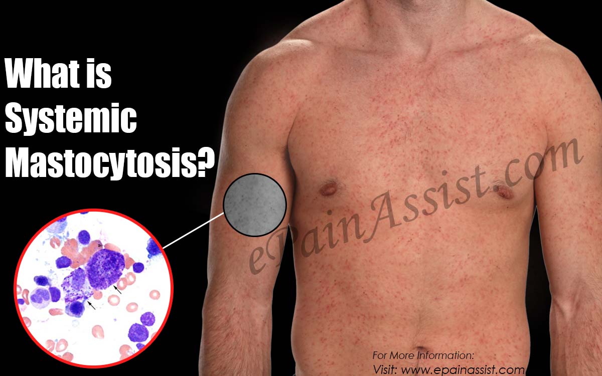
Imagine a 42-year-old woman experiencing episodic flushing. Facial redness. Sudden. Unpredictable. Flushing lasting minutes hours. Associated symptoms. Pruritus itching intense. Facial. Scalp. Tachycardia heart racing. Palpitations. Diarrhea abdominal cramping. Episodes triggered variably. Foods spicy. Alcohol consumption. Stress emotional. Temperature changes. Heat exposure. Medications aspirin NSAIDs. Triggers elusive inconsistent. Symptoms disabling. Occurring unexpectedly. Embarrassing. Work presentations. Meetings. Social settings. Avoided often. Depression anxiety. Possible. She seeks physician evaluation. Multiple physicians. Consultations. Workup. Labs normal. Imaging normal. Carcinoid syndrome. Considered ruled. Allergy component. Antihistamine therapy. Prescribed. Benefit minimal. Years passing. Symptoms persistent. Frustration growing. She mentions skin. Lesions. Tan brown macules small. Scattered. Trunk. Extremities. Urticaria pigmentosa appearance characteristic. Dermographic urticaria lesions. Stroking. Red welts. Appearing. Skin biopsy performed. Mast cell infiltration. Demonstrated. CD117 positive. C-KIT positive. Confirmed. Mastocytosis cutaneous. Suspected. Serum tryptase. Measured. Elevated markedly. Baseline approximately 50 ng/mL. Normal less than 11. Systemic mastocytosis. Diagnosed likely. Bone marrow biopsy. Performed. Mast cell aggregates. Spindle-shaped. Demonstrated. KIT mutation D816V. Detected. Systemic mastocytosis confirmed. Indolent type. Prognosis relatively. Good. Treatment initiated. H1 H2 receptor antagonists. Histamine antagonism. First-line. Cetirizine H1 receptor. Famotidine H2 receptor. Dosing. Regular. Flushing episodes. Reduced frequency. Severity diminished. Cromolyn sodium mast cell. Stabilization. Added. Granule release. Inhibition. Leukotrienes. Prostaglandins. Reduction. Leukotriene. Receptor antagonist. Montelukast. Added. GI symptoms. Abdominal. Diarrhea. Improvement. Possible. Triggers avoided. Aspirin NSAIDs. Avoided avoided absolutely. Anaphylaxis risk. Elevated. Acetaminophen used. Pain relief. Instead. Epinephrine auto-injector. Prescribed. Emergency availability. Critical. Anaphylaxis risk. Elevated. Tryptase levels elevated baseline. She responds well. Symptoms substantially. Reduced. Function improved. Work returned. Social activities. Resumed. Disability. Minimized. She lives managing. Mastocytosis. Regular monitoring. Tryptase levels periodic. Imaging periodic. Leukemic transformation. Surveillance. Understanding systemic mastocytosis enables recognition of this rare mast cell disorder and appropriate management extending survival and improving quality of life. Systemic mastocytosis is a rare hematologic disorder characterized by abnormal proliferation and accumulation of mast cells in multiple organ systems with variable clinical presentations. Systemic mastocytosis accounts for approximately 1 in 150,000 to 1 in 300,000 births. Approximately 1,500 to 3,000 people in United States with mastocytosis. Approximately 10,000 worldwide estimated. Peak onset. Age 40 to 60 years. Adult-onset. Children adolescent-onset possible. Childhood SM rare. Variants multiple subtypes. Indolent systemic. Most common. Smoldering. Aggressive. Mast cell leukemia rare. What makes systemic mastocytosis important to understand is recognizing that while indolent disease is compatible with normal lifespan, aggressive disease poses transformation risk and requires intensive monitoring and therapy. Early diagnosis enables appropriate management preventing complications and improving prognosis. Understanding systemic mastocytosis enables recognition and management. In this comprehensive article, we will explore what systemic mastocytosis is, understand mast cell biology and activation, recognize variable clinical presentations, explore diagnostic criteria and biomarkers, and discover management strategies enabling symptom control and complication prevention.
Understanding Mast Cells and Systemic Mastocytosis Pathophysiology
Before we explore systemic mastocytosis, we need to understand mast cell biology and how abnormal proliferation causes disease. Mast cell development. Origin bone marrow. Hematopoietic progenitor cells. CD34+ cells. Migration tissue peripheral. Differentiation maturation. Tissue environment. Cytokine stimulation. Stem cell factor. SCF. KIT receptor. Signaling. Critical. Mast cell. Development. Maturation. Proliferation. Facilitated. Tissue localization. Connective tissue. Mucous membranes. Around blood vessels. Parasitic. Infection sites. Distribution strategic. Immune surveillance. Facilitated. Mast cell ultrastructure. Granules abundant. Cytoplasmic. Filled mediators. Bioactive. Histamine. Prominent. Tryptase. Serine protease. Chymase. Carboxypeptidase A. Heparin proteoglycan. Others. Granule contents. Preformed. Degranulation. Immediate release. Upon activation. Mediators newly synthesized. Arachidonic acid pathway. Conversion. Prostaglandin D2. PGD2. Leukotriene C4. LTC4. Leukotriene D4. LTD4. Leukotrienes. Vasodilation. Smooth muscle contraction. Inflammation. Mediate. Histamine effects. H1 H2 H3 H4 receptors. Target tissues. H1 receptor. Vascular. Activation endothelial. Increased permeability. Vasodilation. Smooth muscle contraction. Pruritus itching. Mediated. H2 receptor. Gastric acid secretion. Stimulation. Immune response modulation. Tryptase effects. Protease-activated receptors. PAR. Activation. Inflammation tissue remodeling. Facilitation. Increased vascular. Permeability. Smooth muscle contraction. Assisted. Immune response. Modulation. Mast cell activation. Mechanisms multiple. IgE cross-linking. FcεRI high-affinity. IgE receptor. Ligand binding. Receptor oligomerization. Receptor aggregation. Tyrosine kinase activation. Calcium influx. Intracellular. Degranulation. Exocytosis. Activation direct. Complement activation. C3a C5a anaphylatoxins. Receptor stimulation. C5aR. Degranulation mast cell. Triggered. Physical stimuli. Mechanical. Pressure temperature. Vibration. Mastocytosis. Activation. Triggers. Drug induced. Opioids radiocontrast. Hyperosmolar solutions. Vancomycin. Others. Degranulation possible mast cell. Neuropeptide stimulation. Substance P. Nerve fibers. CGRP. Calcitonin gene-related peptide. Stimulation. Mast cell activation. Possible. Pathogen-associated. Molecular patterns. PAMPs. Toll-like receptors. TLR. Mast cell. Activation. Possible. Systemic mastocytosis development. Mast cell proliferation abnormal. Clonal expansion. Somatic mutations. Acquired typically. Germline rare. KIT gene. Chromosome 4. Band q12. Mutations. Approximately 80 to 95 percent. Systemic mastocytosis. D816V substitution mutation. Approximately 80 percent. KIT mutations. Point mutation. Missense. Tyrosine kinase domain. KIT proto-oncogene. Mutant KIT. Constitutive activation. Ligand independent. SCF stimulation. Unnecessary. Signal transduction. Continuous. Proliferation mast cell. Dysregulated. Apoptosis resistance. Anti-apoptotic signals. Enhanced. Mast cell survival. Increased. Clonal proliferation. Progressive. Mast cell burden. Increasing. Tissue infiltration. Multi-organ. Accumulation. Consequences. KIT mutation. Only. Sufficient. Systemic mastocytosis. Development. Additional genetic. Somatic mutations. Present. ASXL1. TET2. SRSF2. TP53. Mutations. Associated. Smoldering aggressive. Systemic mastocytosis. Advanced disease. Associated. Leukemic transformation. Risk. TP53 mutations. Associated. Poor prognosis. Rapidly progressive disease. Pathophysiology mechanisms. Mast cell accumulation tissue. Local inflammation chronic. Mediator release. Excessive. Mast cell infiltration bone marrow. Normal hematopoiesis. Suppression. Cytopenias. Anemia. Thrombocytopenia. Leukopenia. Possible. Mast cell infiltration GI tract. Malabsorption motility disorder. Diarrhea. Possible. Infiltration liver spleen. Hepatosplenomegaly. Possible. Ascites. Possible. Infiltration bone. Lytic lesions. Sclerosis. Osteoporosis. Possible. Pathological fractures. Risk. Infiltration nervous. Central peripheral. Neurologic. Manifestations. Possible. Mast cell mediator effects. Systemic vascular. Histamine release. Hypotension. Shock. Possible severe. Anaphylaxis. Tachycardia compensatory. Cardiac. Chronotropic effects. Arrhythmias. Possible. GI smooth muscle. Contraction. Abdominal cramping. Diarrhea. Possible leukotriene involvement. Respiratory. Smooth muscle bronchi. Contraction. Bronchospasm. Possible. Histamine bronchial. Mucosa stimulation. Edema mucosal. Possible airway. Obstruction. Pruritus. Histamine. Skin mast cell. Receptor H1. Stimulation. Urticaria pigmentosa skin. Lesions mast cell. Infiltration. Visible. Associated pruritus. Typically. Systemic manifestations. Flushing facial. Neck. Trunk. Drenching. Prostaglandin PGD2. Tryptase. Vasodilation. Mediated. The pathophysiology explains how KIT mutations cause clonal mast cell proliferation and excessive mediator release resulting in variable multisystem manifestations.
What is Systemic Mastocytosis?
Systemic mastocytosis is a rare hematologic disorder characterized by clonal mast cell proliferation with abnormal accumulation in bone marrow and other organ systems resulting in variable clinical manifestations and prognosis dependent on disease subtype and burden. Definition. Clonal mast cell disorder. Bone marrow involvement. Diagnostic criterion. Additional organ. Involvement. Typical skin cutaneous. Systemic manifestations. Variable. Mediator-related symptoms predominant often. Neoplastic transformation risk possible. Advanced disease progressive. Prognosis. Subtype dependent. Variants multiple subtypes. Indolent systemic mastocytosis ISM. Bone marrow mast cell burden less than 30 percent. Low disease burden typically. C-findings absent. CRAF signs. Cytopenias organomegaly. Dysplasia. Absence diagnostic. Prognosis favorable. Smoldering systemic mastocytosis SSM. Intermediate disease burden. C-findings present. Risk transformation. Advanced disease. Intermediate. Aggressive systemic mastocytosis ASM. High mast cell burden. Bone marrow greater than 30 percent. C-findings present. Organomegaly marked. Dysplasia cytopenias severe. Prognosis poor. Rapid progression. Transformation risk. High. Mast cell leukemia MCL. Mast cell blasts. Bone marrow greater than 20 percent. Peripheral blood circulating. Mast cells increased. Leukemia acute. Aggressive rapidly progressive. Prognosis. Poor median survival. Months. Cutaneous mastocytosis CM. Skin involvement. Systemic. Bone marrow. No documented. Urticaria pigmentosa UP. Most common cutaneous. Tan brown macules. Skin infiltrated mast cells. Diffuse. Dermographic urticaria. Lesions stroking. Red welts. Appearing minutes. Prognosis skin-limited. Excellent. Systemic manifestations absent typically. Transformation systemic. Risk rare. Prognosis overall. Mastocytosis. Subtypes. Variant mixed. Other rare. Clinical manifestations. Indolent systemic. Manifestations mediator-related. Flushing facial. Pruritus itching. Tachycardia palpitations. GI symptoms diarrhea. Abdominal pain. Possible. Anaphylaxis episodes. Possible. Life-threatening. Risk. Elevated. Triggers variable. Foods. Medications. Stress. Temperature. Individual. Bone marrow infiltration minimal. Cytopenias. Absent typically. Organomegaly absent. Prognosis excellent. Median survival. Normal. Decades. Smoldering systemic. Manifestations similar. Indolent. Severity increased possible. Burden disease. Increased. C-findings present possible. Cytopenias. Mild. Organomegaly. Mild moderate. Prognosis intermediate. Survival years. Median. Variable. Transformation. Risk. Increased. Aggressive systemic. Manifestations. Severe. Burden disease high. C-findings prominent present. Cytopenias marked. Anemia. Thrombocytopenia. Severe. Organomegaly marked. Hepatomegaly splenomegaly massive. Ascites. Possible. Constitutional symptoms fever weight loss fatigue. Associated. Prognosis poor. Median survival years. 5-10. Transformation. Likely. Mast cell leukemia. Manifestations severe aggressive. Peripheral blood. Mast cells circulating. Increased. Constitutional. Symptoms fever. Weight loss. Night sweats. Fatigue. Marked. Cytopenias severe. Hemorrhage infection. Risk. High. Organomegaly marked. Infiltration hepatic. Splenic. Bone marrow. The clinical features reflect clonal mast cell burden with variable distribution and mediator release intensity determining prognosis from excellent indolent to poor aggressive leukemic disease.
Recognizing Systemic Mastocytosis: Clinical Presentations and Diagnostic Challenges
Systemic mastocytosis has variable presentations recognizable by mediator symptoms, cutaneous lesions, and variable organ involvement often initially misdiagnosed as allergy, carcinoid, or psychiatric disease. Flushing pruritus presentation (indolent). Middle-aged adult. Age 40 to 60 years. Flushing episodes facial. Recurrent. Sudden. Unpredictable. Flushing lasting minutes to hours. Severe flushing drenching. Associated. Pruritus intense itching. Localized generalized. Facial. Scalp. Trunk. Urticaria possible hives visible. Tachycardia palpitations heart racing. Associated. Dizziness lightheadedness. Possible syncope. Rare. Diarrhea abdominal cramping. GI symptoms associated. Frequency episodes variable. Daily. Weekly. Monthly. Unpredictable. Triggers identified. Spicy foods. Alcohol. Histamine foods. Aged cheese. Cured meats. Fermented foods. Stress emotional. Temperature. Heat exposure. Cold exposure. Medications triggering. Aspirin. NSAIDs. Opioids radiocontrast. Vancomycin. Anesthesia induction. Others. Triggers variable. Inconsistent. Symptoms unpredictable. Impact. Psychological significant. Anxiety. Depression. Avoidance. Social activities. Occupation. Symptoms. Physical examination. Skin findings. Urticaria pigmentosa. Characteristic. Tan brown macules. Hyperpigmented. Trunk. Extremities. Scattered. Dermographic urticaria. Positive. Lesions stroking. Red. Welts. Minutes. Appearing. Urticaria pigmentosa skin. Finding. Visible. Associated. Pruritus. Vital signs. Tachycardia possibly. Blood pressure. Slightly elevated possible. Labs. Serum tryptase. Elevated. Baseline approximately 20-50 ng/mL. Normal less than 11. Diagnostic significance. Bone marrow biopsy. Mast cells. Aggregates spindle-shaped. Demonstrated. CD117 positive. CD13 CD33. Possible positive. KIT mutation D816V. Detected. Systemic mastocytosis confirmed. Imaging. Abdominal imaging. Hepatosplenomegaly. Absent. Organomegaly absent. DEXA scan. Bone density. Osteoporosis possible. Systemic mastocytosis. Indolent type. Diagnosed. Urticaria pigmentosa skin presentation. Child adolescent. Age 5 to 20 years. Skin lesions. Tan brown macules. Pigmented. Trunk. Extremities. Present since childhood. Dermographic urticaria lesions. Stroking. Red welts. Appearing. Pruritus. Minor itching. Cosmetic concern. Primary motivation. Evaluation. Constitutional symptoms. Absent. GI symptoms. Absent. Anaphylaxis episodes. Absent. Labs. Serum tryptase. Normal or minimally elevated. Less than 20 ng/mL. Bone marrow biopsy. Not performed. Cutaneous mastocytosis. Diagnosed clinically. Skin biopsy. Confirmation. Mast cells skin. Infiltration. CD117 positive. Demonstrated. Prognosis excellent. Skin-limited. Systemic. Progression. Rare. Observation monitoring. Sufficient. No treatment. Necessary. Cosmetic. Tolerance improved. Adulthood. Prognosis excellent. Normal lifespan expected. GI manifestations presentation. Adult. Age 30 to 50 years. Diarrhea chronic. Persistent. Months years. Alternating. Constipation possible. Abdominal pain. Cramps. Associated. Bloating. Flatulence. Malabsorption. Possible. Weight loss. Progressive. Nutrient deficiencies. Possible. Fatigue weakness. Associated. IBS irritable bowel. Syndrome. Diagnosed initially. Dietary modifications. Attempted. Benefit minimal. GI medications. Antidiarrheals. Loperamide. Tried. Minimal benefit. Flushing episodes. Associated. Tachycardia. Palpitations. GI manifestations. Temporal relationship. Episodes clustered. Triggering mediator. Suspected. Labs. Serum tryptase. Mildly elevated. Approximately 15-25 ng/mL. Borderline. Bone marrow biopsy. Performed. Mast cell infiltration. Demonstrated. KIT mutation. Detected. Systemic mastocytosis. Diagnosed. Mast cell burden. Mild. Indolent expected. Management. Cromolyn sodium. Mast cell stabilization. Diarrhea reduction. Possible. H1 H2 antagonists. Added. Mediator blockade. GI symptoms. Improvement. Possible. Symptoms substantially. Reduced. Leukotriene receptor. Antagonist montelukast. Added. Abdominal symptoms. Improvement. Continued. Anaphylaxis presentation (emergency). Young adult. Age 20 to 40 years. Anaphylaxis episode sudden. Severe. Facial flushing. Urticaria widespread. Pruritus intense. Angioedema throat lips. Airway compromise risk. Stridor. Respiratory distress. Wheezing bronchospasm. Cardiovascular. Collapse hypotension. Tachycardia. Syncope loss consciousness. GI abdominal. Pain diarrhea. Vomiting. Emergency treatment. Epinephrine IM 0.3-0.5 mg. Administered. IV fluids. Resuscitation. Provided. Antihistamines H1 H2. IV corticosteroids. Given. Hospital admission. Monitoring. Intensive. Recovery gradual. Trigger identification. Difficult. Anaphylaxis. Unexplained. Recurrent episodes possible. Risk. Elevated. History mastocytosis family. Possible. Labs. Serum tryptase. Markedly elevated. Acute episode. During possible 100-200 ng/mL. Baseline. Elevated. 20-50 ng/mL. Persistent elevation. Systemic mastocytosis. Suspected. Bone marrow biopsy. Mast cells increased. KIT mutation. Systemic mastocytosis. Confirmed. Anaphylaxis recurrent. Risk. Epinephrine auto-injectors. Prescribed. Multiple. Availability. Critical. Emergency preparedness. Essential. Advanced disease presentation (aggressive smoldering). Adult. Age 50 to 70 years. Constitutional symptoms fever. Weight loss progressive. Night sweats. Fatigue debilitating. Cytopenias anemia. Thrombocytopenia. Bleeding bruising. Infection risk. Elevated. Organomegaly hepatosplenomegaly marked. Abdominal distension ascites. Possible. Skin. Manifestations cutaneous. Possible or absent. GI symptoms prominent abdominal. Pain diarrhea. Labs. Serum tryptase. Markedly elevated 50-100+ ng/mL. Bone marrow. Mast cells increased significantly. Burden. Greater than 10-30 percent. Possible. Dysplasia morphologic. Atypical cells. Possible. KIT mutation D816V. Detected. Additional mutations. ASXL1. TET2. SRSF2. Possible. Aggressive systemic. Mastocytosis diagnosed. Or smoldering. C-findings present. Risk transformation. Elevated. Chemotherapy cytoreduction. Consideration. Discussed. Prognosis. Poor. Survival years. Median 5-10. Advanced disease.
The diverse presentations require high clinical suspicion and appropriate diagnostic evaluation for recognition particularly anaphylaxis episodes with unexplained mediator symptoms.
Diagnosis: Diagnostic Criteria and Biomarkers
Diagnosing systemic mastocytosis requires bone marrow biopsy demonstrating mast cell aggregates with KIT mutation confirmation combined with clinical criteria and serum tryptase elevation. WHO diagnostic criteria. Bone marrow biopsy. Mast cells aggregates spindle-shaped. Dense multifocal. Infiltrates demonstrated. Criterion major. One additional. Criterion minor. Diagnosis systemic. Mastocytosis. Confirmed. Minor criteria. KIT mutation D816V bone marrow. Detected. Mast cell tryptase serum. Baseline greater than 20 ng/mL. Bone marrow biopsy. Mast cells greater than 25 percent. CD2 CD25. Mast cells. Abnormal expression detected. Criterion major satisfied. Bone marrow aggregates. Spindle-shaped mast cells morphology characteristic. Dense infiltrates. Multifocal. Greater than 15 cells. Aggregates. CD117 positive immunohistochemistry. Mast cell marker. CD13 CD33 coexpression. Possible abnormal phenotype. Suggested. Serum tryptase baseline. Measured. Normal less than 11 ng/mL. Elevated greater than 11 ng/mL. Significant. Greater than 20 ng/mL. Markedly. Elevated. Diagnostic. Tryptase elevation acute. Episode. During anaphylaxis. Episode. Acute tryptase. Peak. Greater than baseline. Approximately 1.2 times. Acute episode. Diagnostic. Tryptase baseline. Persistent. Elevated. Bone marrow. Involvement suggested. Genetic testing. KIT mutation. D816V typically. Approximately 80-95 percent systemic. Mastocytosis. Detected. Testing. Bone marrow aspirate. DNA extracted. PCR targeted. KIT D816V mutation. Detection. Sequencing direct. Possible. Sanger sequencing. Standard. Next-generation sequencing. NGS. Increasingly. Used. KIT wild-type. Systemic mastocytosis. Possible. KIT mutation. Negative. Alternative genes. PDGFRA. Mutations. Possible rare. Seronegative systemic. Mastocytosis. Rare. PDGFRA mutations. PDGFRA. Platelet-derived growth. Factor receptor alpha. Gene. Mutations possible. Rare. Tyrosine kinase inhibition. Sensitive. Imatinib. Possible therapy. Platelet derived growth. Factor receptor. Inhibitor imatinib. Benefit. Possible. Other somatic mutations. Advanced disease. ASXL1. TET2. SRSF2. TP53. Others. Detected. NGS mutation panel. Multigen. Advanced systemic. Mastocytosis. Aggressive smoldering. Identified. Prognosis. Prognostic implications. Mutation burden. Assessment. Diagnostic algorithm. Middle-aged adult. Flushing pruritus. Tachycardia episodes. Cutaneous lesions. Urticaria pigmentosa. Bone marrow biopsy. Performed. Mast cell aggregates. Spindle-shaped. CD117 positive demonstrated. Criterion major. Serum tryptase. Elevated. Greater than 20 ng/mL. Criterion minor. Genetic testing. KIT D816V mutation. Detected. Criterion minor. Systemic mastocytosis diagnosis. Confirmed. Indolent type. Established probable. Clinical features compatible. Prognosis discussed. Management planned. Imaging. Abdominal. CT. Hepatosplenomegaly. Assessment. DEXA scan. Bone density. Osteoporosis. Assessment. Baseline established. Monitoring. Serial. The diagnosis requires bone marrow biopsy demonstrating mast cell aggregates with genetic confirmation enabling subtype classification and prognosis determination.
Management: Mediator-Blocking Therapy and Targeted Treatment
Systemic mastocytosis management focuses on mediator antagonism controlling symptoms, targeted therapy for KIT mutations, and monitoring for disease progression or transformation. Mediator antagonists H1 H2 receptor. First-line therapy. H1 receptor antagonists. Cetirizine fexofenadine levocetirizine. Non-sedating. Preferred. Diphenhydramine. Sedating. Alternative. Dosing. Regular scheduled. Prophylactic. As-needed dosing insufficient often. Twice daily dosing. Typical. H2 receptor antagonists. Famotidine ranitidine. Gastric acid. Secretion reduction. Gastric. Mucosal protection. Histamine H2. Receptor effects. Antagonism. GI manifestations. Improvement. Combined H1 H2. Blockade. Mediator antagonism complete. Superior single agent. Benefit combination. Demonstrated. Cromolyn sodium mast cell. Stabilization. Mechanism. Mast cell. Granule release prevention. Mediator preformed. Blocking. Dosing 100-200 mg. Orally. Three times daily. Consistent. Dosing. Regular maintenance. Importance. Benefit onset. Gradual weeks possible. Compliance sustained. Important. Efficacy variable. Some. Patients. Excellent response. Others. Limited. Trial therapy. Necessary. Leukotriene receptor antagonists. Montelukast zafirlukast. Leukotriene C4 D4. Receptor blockade. GI smooth muscle. Contraction reduction. Diarrhea. Abdominal cramping. Improvement. Asthma concurrent. Bronchospasm. Benefit possible. Dosing montelukast. 10 mg nightly. Typical. Efficacy variable. Trial needed. Gastric acid suppression. PPI proton pump inhibitor. Omeprazole pantoprazole. Gastric. Acid reduction. Mastocytosis. GI involvement. Diarrhea severe. Associated. Acid suppression. Benefit. Possible. H2 antagonist. Combined. Efficacy enhanced. Possible. Targeted therapy tyrosine kinase inhibitors. TKI. Imatinib. KIT proto-oncogene inhibitor. D816V mutation. Typically resistant. Standard imatinib dosing. Sensitivity variable. Higher dosing. Possible trial. Increased toxicity. Risk. Limited benefit. Some patients. Dasatinib. Alternative TKI. D816V mutation. Sensitivity possible. Clinical utility limited. Midostaurin. FLT3 inhibitor. KIT inhibition activity. Possible. Aggressive advanced systemic. Mastocytosis. Studied. Efficacy. Demonstrated. Clinical utility. Emerging. Avapritinib. Novel KIT D816V-directed. Inhibitor. Recently developed. Advanced aggressive. Systemic mastocytosis. Studied. Efficacy encouraging. Early trial results. Approval. FDA possible pending. Anaphylaxis management. Emergency treatment. Epinephrine IM. 0.3-0.5 mg. Intramuscular injection. Thigh IM. Optimal. Rapid absorption. Vasoconstriction cardiovascular. Support. Epinephrine auto-injector. Prescribed. Multiple. Home. Availability. Critical. Emergency preparedness. Written action plan. Provided. Antihistamines IV. H1 H2 antagonists IV. Following epinephrine. Complementary therapy. Corticosteroids IV. Methylprednisolone hydrocortisone. Inflammation reduction. Following epinephrine. IV fluid resuscitation. Hypotension treatment. Vasopressors. Norepinephrine dopamine. Possible. Shock refractory. Epinephrine. Observation hospitalization prolonged. Anaphylaxis episode. Monitoring. Biphasic reaction. Recurrent. Possible. Time observation. Adequate. Trigger identification avoidance. Triggers variable. Identified when possible. Avoided diligently. Oral epinephrine. Investigational. Sublingual. Absorption improved possible. Auto-injector epinephrine. Current standard. Counseling. Trigger avoidance. Foods triggering. Identified. Avoidance. Strict. Medications triggering aspirin. NSAIDs. Opioids radiocontrast. Avoidance. Absolute. Acetaminophen. Pain relief alternative. Anesthesia anesthesiologist. Informed. Mastocytosis diagnosis. Anesthesia triggering. Possible drugs. Identification. Avoidance. Pre-operative anesthesia. Planning. Coordination. Important. Symptom monitoring. Keeping symptom diary. Triggers. Symptoms. Temporal relationships. Identification. Possible. Physician. Review. Education patient. Trigger avoidance. Effective. Quality life. Improved. Significantly. Bone marrow monitoring. Indolent systemic. Mastocytosis. Stable often. Monitoring periodic. Serum tryptase. Annual. Imaging CT. Annual. Reassess. Disease progression surveillance. Smoldering aggressive systemic. Mastocytosis. Monitoring more frequent. Tryptase every 3-6 months. Imaging every 6-12 months. Bone marrow biopsy. Periodic. Transformation surveillance. Leukemic transformation. Risk increased. Smoldering aggressive. Advanced disease. Blast percentage bone marrow. Monitoring. Peripheral blood. Mast cells. Monitoring. Systemic symptoms development. Constitutional. Cytopenias progressive. Fever. Rapid deterioration. Leukemic transformation. Suggested. Bone marrow biopsy repeat. Immediately. Aggressive treatment chemotherapy. Cytoreduction. Indicated. Cytoreduction chemotherapy. Advanced disease. Hypomethylating agents. Azacitidine decitabine. Possible trial. Aggressive systemic. Mastocytosis. Efficacy limited. Responses partial. Chemotherapy conventional. Cladribine pentostatin. Possible. Responses possible. Toxicity. Significant. Clinical trial enrollment. Consideration. Novel therapies. Possible options. Supportive care. Osteoporosis management. Bone involvement. Systemic mastocytosis. Osteoporosis. Risk. DEXA scan. Baseline. Periodic. Monitoring. Calcium vitamin D supplementation. Bone health support. Bisphosphonate therapy. Possible. Bone loss. Significant. Fracture prevention. Important. Anemia. Iron supplementation. B12 folate. Deficiency. Corrected. Transfusion. Possible. Hemoglobin. Severely reduced. Bleeding. Risk. Transfusion threshold. 7-8 g/dL typical. Higher. Symptoms. Compromise oxygen. Tissue. Thrombocytopenia. Platelet transfusion. Possible. Bleeding. Severe risk. Platelet count. Very low. Infection. Prophylaxis possible. G-CSF granulocyte colony-stimulating factor. Possible. Neutropenia. Severe. Infection. Risk. Molecular monitoring. KIT D816V mutation. Burden. Monitoring possible research. Protocol. Clinical trial. Novel biomarkers. Emerging. Mast cell burden assessment. New method. Therapy response monitoring. Possible. The comprehensive approach addresses mediator antagonism with H1 H2 antagonists and cromolyn, targeted therapy for KIT mutations, and monitoring for disease progression or transformation.
Frequently Asked Questions (FAQs)
Q1: Is systemic mastocytosis life-threatening?
Depends subtype severity. Indolent systemic. Mastocytosis. Life expectancy normal. Lifespan typical. Complications. Rare serious. Anaphylaxis episodes. Risk. Manageable epinephrine. Preparedness education. Smoldering aggressive systemic. Mastocytosis. Life expectancy reduced. Median survival years 5-10 approximate. Advanced disease. Progression. Transformation risk. Increased. Mast cell leukemia. Life-threatening acutely. Prognosis poor. Median survival months. Chemotherapy cytoreduction. Possible but uncertain benefit. Indolent type long-term outlook. Excellent generally. Transformation. Risk low. Most patients. Die other. Causes. Age-related.
Q2: How is systemic mastocytosis inherited?
Genetic disorder somatic. Acquired mutations. Clonal. Bone marrow cell. Affected. Mutation acquired. Inherited not typically. Familial clustering rare. Hereditary alpha-tryptasemia HAT. Germline. Polymorphism TPSAB1 gene. Tryptase alpha gene duplication. Familial possible. Baseline tryptase elevation. Constitutional. Systemic mastocytosis. Risk. Increased possible. Hereditary mastocytosis familial. Extremely rare. Germline KIT mutation. PDGFRA. Possible. Genetic counseling offered. Familial cases. Relatives. Screening possible. Genetic testing. Limited utility. Sporadic disease. Relatives not. At genetic risk typically.
Q3: Can I be cured?
Cure unlikely. Genetic disease clonal. Therapy symptomatic goal. Current mediator antagonism. Targeted therapy KIT inhibitors. Disease stabilization possible sustained. Indolent type stable. Often years decades. Smoldering aggressive transformation. Risk progression. Advanced disease chemotherapy. Cytoreduction goal. Leukemia remission cure. Possible but unlikely durable. Gene therapy investigation. Future possibly. Mutation-specific correction. Possible future. Cure development timeline. Uncertain. Years decades possible. Current therapy. Management symptom control symptom control. Prognosis variable. Subtype specific.
Q4: What triggers anaphylaxis?
Variable triggers. Individual. Common. Foods spicy. Alcohol. Histamine foods aged. Cheese. Cured meats. Fermented foods. Medications aspirin. NSAIDs. Opioids. Radiocontrast. Vancomycin. Others. Stress emotional. Temperature extremes. Heat cold. Infection. Possible trigger. Idiopathic anaphylaxis no. Trigger identified. Possible. Trigger identification. Diary keeping. Helpful. Avoidance strict. Trigger-identified. Important. Triggers unknown variable. Unpredictability. Anxiety. Stress related. Epinephrine auto-injector emergency. Availability. Critical. Preparedness education. Essential.
Q5: Will I need a bone marrow transplant?
Unlikely indolent. Systemic mastocytosis. Bone marrow transplant. Not indicated. Stable disease. Management medical sufficient. Advanced disease. Aggressive transformation leukemic. Bone marrow transplant. Consideration possible. Clinical trial. Possible. Therapy targeted chemotherapy. Cytoreduction. First-line. Response inadequate. Bone marrow transplant transplantation. Possible consideration. Allogeneic transplant. Donor matched. HLA. Risks substantial. Graft-versus-host disease. GVHD. Mortality perioperative. Morbidity. Risk-benefit assessment. Critical. Transplant. Specialist. Hematology-oncology. Referral. Discussion. Therapy options. Available.
Key Takeaways
Systemic mastocytosis is rare hematologic clonal mast cell disorder abnormal proliferation. Approximately 1 in 150,000-300,000 births. Approximately 1,500-3,000 United States. Peak onset age 40-60 years. Multiple subtypes indolent smoldering aggressive mast cell leukemia. Indolent most common. Vascular mast cell leukemia rare life-threatening. Pathophysiology. KIT gene mutations D816V approximately 80-95 percent systemic mastocytosis. Constitutive activation tyrosine kinase. Ligand-independent proliferation. Clonal mast cell expansion. Bone marrow involvement diagnostic. Multisystem involvement variable. Mediator excessive release histamine tryptase leukotrienes prostaglandins. Systemic manifestations. Clinical features. Flushing pruritus tachycardia palpitations GI diarrhea abdominal pain anaphylaxis episodes life-threatening. Urticaria pigmentosa cutaneous skin lesions characteristic tan-brown macules dermographic urticaria. Organomegaly hepatosplenomegaly possible advanced disease. Constitutional symptoms fever weight loss advanced disease. Diagnosis. Bone marrow biopsy major criterion mast cell aggregates spindle-shaped multifocal CD117 positive. Minor criteria KIT mutation D816V serum tryptase baseline elevated >20 ng/mL mast cells >25 percent bone marrow CD2 CD25 abnormal expression. WHO criteria diagnosis confirmed major one additional minor criteria. Serum tryptase elevated baseline > 11 ng/mL diagnostic significant > 20 ng/mL. Genetic testing KIT mutation D816V confirmed detection. Management. H1 H2 receptor antagonists histamine mediator blockade cetirizine famotidine first-line combined therapy. Cromolyn sodium mast cell stabilization granule release inhibition. Leukotriene receptor antagonist montelukast GI symptoms improvement. Anaphylaxis emergency treatment epinephrine IM auto-injector availability critical. H1 H2 antihistamines IV corticosteroids IV fluids resuscitation. Trigger avoidance identification foods aspirin NSAIDs opioids medications. Targeted therapy tyrosine kinase inhibitors KIT mutations imatinib dasatinib midostaurin avapritinib D816V directed. Bone marrow monitoring periodic tryptase imaging transformation surveillance. Prognosis. Indolent systemic mastocytosis excellent normal life expectancy. Smoldering intermediate prognosis transformation risk. Aggressive poor prognosis median 5-10 years survival. Mast cell leukemia poor median months survival. Advanced disease chemotherapy cytoreduction possible bone marrow transplant consideration.
References
- World Health Organization (WHO). “Systemic Mastocytosis: Diagnosis and Management.” Retrieved from https://www.who.int/
- National Institutes of Health. “Mastocytosis Information.” Retrieved from https://www.nih.gov/
- American Academy of Allergy, Asthma and Immunology (AAAAI). “Mastocytosis Resources.” Retrieved from https://www.aaaai.org/
- Mayo Clinic. “Systemic Mastocytosis: Diagnosis and Treatment.” Retrieved from https://www.mayoclinic.org/
- National Organization for Rare Disorders (NORD). “Systemic Mastocytosis.” Retrieved from https://rarediseases.org/
- American Society of Hematology. “Mastocytosis Guidelines.” Retrieved from https://www.hematology.org/
Related Articles on ObserverVoice.com
Explore more health and science topics on our platform:
- Mast Cell Disorders: Understanding Immune Cell Dysfunction
- Anaphylaxis: Understanding Severe Allergic Reactions
- Hematologic Malignancies: Understanding Blood Cell Cancers
- Genetic Testing: Understanding DNA-Based Diagnosis
- Tyrosine Kinase Inhibitors: Understanding Targeted Cancer Therapy
- Bone Marrow Biopsy: Understanding Diagnostic Procedure and Interpretation
Disclaimer
This article provides educational information adapted from publicly available health sources including WHO materials. This content is for informational and educational purposes only and does not constitute medical advice, diagnosis, or treatment recommendations. [ObserverVoice.com] is a news and information platform—not a healthcare provider. If you experience recurrent flushing, pruritus, tachycardia episodes with variable triggers, cutaneous lesions with urticaria pigmentosa appearance, or unexplained anaphylaxis episodes, consult physicians for evaluation. Systemic mastocytosis diagnosis requires bone marrow biopsy demonstrating mast cell aggregates (greater than 15 cells per aggregate, spindle-shaped morphology, CD117 positive) combined with diagnostic criteria (major criterion bone marrow aggregates plus one minor criterion: KIT D816V mutation, serum tryptase baseline >20 ng/mL, mast cells >25 percent bone marrow, or CD2/CD25 abnormal mast cell expression). Serum tryptase baseline elevation (>11 ng/mL) is significant; markedly elevated (>20 ng/mL) is diagnostic. Genetic testing confirming KIT D816V mutation is important for prognosis and therapy. Management includes H1 and H2 receptor antagonists (cetirizine, famotidine) as first-line mediator antagonism with combined therapy superior. Cromolyn sodium stabilizes mast cells preventing granule release. Leukotriene receptor antagonists (montelukast) manage GI manifestations. Targeted tyrosine kinase inhibitors (imatinib, midostaurin, avapritinib) inhibit KIT mutations. Anaphylaxis episodes require emergency epinephrine (0.3-0.5 mg IM) with auto-injectors prescribed for home availability. Disease subtype determines prognosis: indolent systemic mastocytosis has excellent prognosis with normal life expectancy while advanced aggressive disease has median survival 5-10 years with transformation risk. With appropriate diagnosis and management, quality of life improves. Always seek guidance from qualified allergists, hematologists, and mastocytosis specialists experienced in comprehensive management.
Observer Voice is the one stop site for National, International news, Sports, Editor’s Choice, Art/culture contents, Quotes and much more. We also cover historical contents. Historical contents includes World History, Indian History, and what happened today. The website also covers Entertainment across the India and World.

