

Primary Biliary Cholangitis (PBC): The Autoimmune Liver Bile Duct Disease

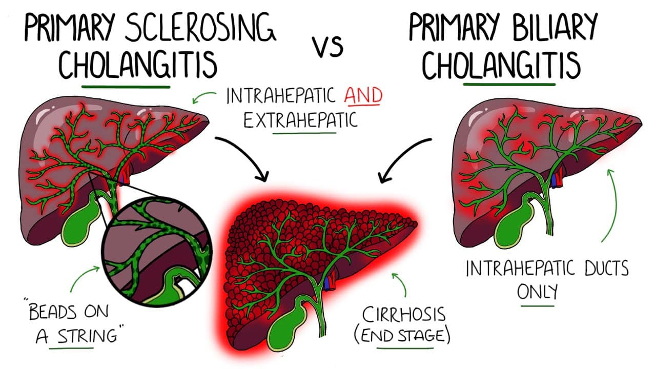
Imagine bile ducts—tiny channels transporting digestive fluid throughout your liver—becoming chronically inflamed and slowly destroyed by your own immune system. As ducts disappear, bile backs up into your liver damaging hepatocytes. Your skin becomes unbearably itchy from bile salt accumulation. Jaundice yellows your skin and eyes. Gradually, your liver scarring progresses toward cirrhosis and liver failure. This is primary biliary cholangitis—a serious autoimmune disease uniquely targeting bile ducts within the liver. Primary biliary cholangitis, commonly abbreviated as PBC, is a chronic autoimmune disease causing progressive inflammation and destruction of intrahepatic bile ducts—the small channels carrying bile within the liver. The immune system mistakenly attacks bile duct epithelial cells causing chronic inflammation. The inflammation gradually destroys bile ducts. As ducts are destroyed, bile cannot flow normally. Bile backs up into the liver and bloodstream. The accumulated bile damages hepatocytes and causes jaundice. Progressive bile duct destruction leads to cirrhosis and liver failure if untreated. Primary biliary cholangitis affects approximately 1 to 2 per million people worldwide, though prevalence is higher in developed countries. The disease is rare compared to other liver diseases but is the most common autoimmune liver disease in many countries. Women comprise approximately 90 percent of PBC patients. The disease predominantly affects middle-aged women, typically developing between ages 40 and 60. Men rarely develop PBC but when they do, disease is typically more severe. What makes primary biliary cholangitis distinctive is the specific autoantibody pattern. Approximately 95 percent of PBC patients have anti-mitochondrial antibodies (AMA), particularly anti-M2 antibodies. This specific autoantibody pattern makes PBC easily recognizable and distinguishes it from other autoimmune liver diseases. Modern treatments have dramatically improved outcomes. Ursodeoxycholic acid, the first-line treatment, slows disease progression and improves liver function in most patients. Newer medications including obeticholic acid and fibrates show promise. With appropriate treatment started early, many PBC patients achieve stable disease and avoid progression to cirrhosis. In this comprehensive article, we will explore what primary biliary cholangitis is, understand how bile duct destruction develops, recognize early warning symptoms, learn about serious complications, understand diagnosis methods, explore available treatments, and discover management strategies for preserving liver function and quality of life.
Understanding Bile Ducts and Normal Bile Function
Before we explore primary biliary cholangitis, we need to understand bile ducts and normal bile function. The liver produces bile—a green-yellow digestive fluid essential for fat digestion and absorption. Bile contains bile acids, cholesterol, bilirubin, and other substances. Hepatocytes continuously produce bile. The bile flows through increasingly larger bile ducts. Small ducts within liver tissue collect bile from hepatocytes. These small ducts merge into progressively larger ducts. The largest duct within the liver is the hepatic duct. The hepatic duct merges with the cystic duct from the gallbladder forming the common bile duct. The common bile duct exits the liver and pancreas, emptying into the small intestine (duodenum). Bile ducts are lined with specialized epithelial cells called cholangiocytes. Cholangiocytes form a protective barrier between bile and the duct wall. Cholangiocytes also modify bile composition. Healthy bile ducts allow normal bile flow. Bile flows continuously into the intestine. After meals, the gallbladder contracts releasing stored bile into the intestine increasing fat digestion. In the intestine, bile acids emulsify fats allowing enzymatic digestion and absorption. Bile acids are reabsorbed in the terminal ileum and recycled back to the liver. This enterohepatic circulation recycles bile acids multiple times daily. Bilirubin in bile comes from hemoglobin breakdown. Hepatocytes take up bilirubin from blood and excrete it into bile. Bilirubin travels through bile ducts to the intestine where it is converted to stercobilin giving stool its brown color. Normal bile flow is crucial for digestion and elimination of bilirubin and cholesterol. Bile duct damage impairs these essential functions. Understanding that bile ducts are essential for bile flow and elimination of bilirubin helps explain why their destruction in PBC causes jaundice and other complications.
What is Primary Biliary Cholangitis?
Primary biliary cholangitis is a chronic autoimmune disease causing progressive inflammation and destruction of intrahepatic bile ducts—the small channels within the liver carrying bile. The disease is characterized by immune attack specifically targeting bile duct epithelial cells. In primary biliary cholangitis, the body’s immune system becomes dysregulated. Autoantibodies—antibodies against the body’s own proteins—attack bile duct epithelial cells. The most characteristic autoantibody is anti-mitochondrial antibody (AMA), particularly anti-M2 antibodies. These antibodies target pyruvate dehydrogenase complex (PDC) in mitochondria—the energy-producing organelles in cells. Although the antibodies are called anti-mitochondrial, they actually attack the antigens on bile duct cells. T lymphocytes and other immune cells infiltrate bile ducts. These immune cells produce inflammatory chemicals destroying bile duct epithelium. The bile ducts are progressively damaged and destroyed. As ducts are destroyed, the normal network of bile channels is lost. Bile cannot flow normally. Bile backs up into remaining ducts. Bile accumulates in hepatocytes. The stagnant bile damages liver cells. Hepatocytes die. The liver attempts repair by producing fibrosis. Progressive fibrosis leads to cirrhosis. The disease follows a predictable progression though the rate varies between individuals. Early stages involve active inflammation of bile ducts with preservation of duct architecture. Intermediate stages involve progressive duct destruction with loss of intrahepatic ducts. Late stages involve extensive fibrosis and cirrhosis. Without treatment, most patients progress from early to late stages over 10 to 20 years. However, with effective treatment, progression is arrested or slowed. What causes the immune system to attack bile ducts is incompletely understood. Genetic factors are important—PBC runs in families. Specific HLA gene types increase susceptibility. However, genetics alone does not cause PBC. Environmental factors are also necessary. Bacterial infections have been suspected as potential triggers. Specific bacteria including E. coli expressing antigens mimicking bile duct antigens might trigger autoimmune response. The molecular mimicry—bacteria antigens resembling bile duct antigens—might activate immune cells attacking both bacteria and bile ducts. Viral infections including hepatitis C virus might trigger PBC development. Xenobiotics—foreign chemical substances—including pesticides and industrial chemicals might trigger PBC. However, the specific environmental trigger, if any, remains unknown. Gender hormones clearly influence PBC development. Women are nine times more likely than men to develop PBC. Estrogen might promote autoimmune response. This hormonal influence is typical of autoimmune diseases generally. PBC is classified into two stages based on histology: nonsymptomatic (asymptomatic) and symptomatic. Approximately 50 percent of patients are asymptomatic at diagnosis, discovered through abnormal liver tests. These patients often have better prognosis than symptomatic patients because disease is diagnosed earlier. Symptomatic patients present with jaundice, itching, or other symptoms indicating more advanced disease.
Recognizing Early Symptoms: Signs of Bile Duct Disease
Primary biliary cholangitis symptoms often develop insidiously, sometimes causing subtle symptoms initially attributed to other causes. Recognizing early symptoms prompts medical evaluation allowing earlier diagnosis and treatment. Itching (pruritus) is the most characteristic and often the most bothersome symptom. Severe itching affects most PBC patients. The itching is caused by accumulation of bile salts in blood. Bile salts deposit in skin causing intense itching. The itching is often worse at night disrupting sleep. The itching is sometimes so severe patients scratch until bleeding. The itching sometimes precedes jaundice by months or years. Some patients have severe itching as the only symptom despite having advanced liver disease. The itching causes significant quality of life impairment. Fatigue is extremely common. Overwhelming exhaustion develops affecting daily functioning. Fatigue is often disproportionate to physical activity. Patients report tiredness affecting work and social activities. Fatigue sometimes precedes jaundice or other obvious symptoms. Jaundice develops as liver function deteriorates. Yellowing of skin and whites of eyes occurs. Jaundice results from elevated bilirubin in blood. Bilirubin accumulates because damaged liver cannot excrete it normally. Jaundice indicates significant liver dysfunction. Pale or clay-colored stools develop from reduced bile flow to intestines. Without bile, stool lacks normal brown coloration. Pale stools indicate impaired bile flow. Dark urine develops from bilirubin excretion. The urine becomes dark brown or cola-colored. Dark urine indicates elevated bilirubin in blood. Abdominal pain or discomfort develops in some patients. The pain is usually localized to upper right abdomen where the liver sits. The pain might be dull or sharp. Abdominal pain suggests hepatic inflammation. Joint and muscle pain develop in some patients. Joints become painful and sometimes swollen. The joint pain sometimes precedes liver symptoms. Arthralgias might be attributed to arthritis before PBC is diagnosed. Dry eyes and dry mouth (Sjögren’s syndrome) develop in some PBC patients. Lacrimal and salivary glands become involved. Approximately 50 percent of PBC patients have coexisting Sjögren’s syndrome. Other autoimmune disease coexistence is common in PBC. Xanthomas develop in some patients. These are yellowish lipid deposits in skin and around eyes. Xanthomas result from elevated cholesterol from impaired bile excretion. Xanthomas around eyes create puffy appearance. Bone pain develops in some patients. Osteoporosis from liver disease and bile salt malabsorption weakens bones. Bone fractures from minor trauma indicate severe osteoporosis. Hepatomegaly (enlarged liver) develops as disease progresses. The liver becomes enlarged from inflammation and scarring. Enlargement might be detected on physical examination. Splenomegaly (enlarged spleen) develops in advanced disease. The spleen enlarges from portal hypertension. Spleen enlargement might be palpable on examination. Ascites (abdominal fluid) develops in advanced disease. Fluid accumulates in abdomen from liver dysfunction and portal hypertension. Abdomen swells and becomes tender. Ascites indicates significant liver damage. Many early PBC patients have no symptoms. The disease is discovered when blood tests ordered for other reasons show abnormal liver function or positive AMA. Asymptomatic patients diagnosed early generally have better outcomes than symptomatic patients.
Understanding Bile Duct Destruction and Complications
Understanding how progressive bile duct destruction develops helps explain why early treatment is crucial. Bile duct injury initiates immune response. The trigger—infection, xenobiotic exposure, or molecular mimicry—causes initial bile duct damage. The damage exposes antigens triggering immune response. Autoantibodies against these antigens develop. Immune cells attack bile duct epithelium. Bile duct inflammation develops. Lymphocytes infiltrate bile duct walls producing inflammatory chemicals. The inflammation damages cholangiocytes. Cholangiocytes die. The dead cells are removed leaving damaged duct. Duct destruction progresses. With continued immune attack, more ducts are damaged. The bile duct network shrinks. Fewer ducts remain to carry bile. Bile flow becomes impaired. Cholestasis develops—bile accumulation in liver. Cholestatic liver disease develops. Bile backs up causing damage. Bilirubin accumulates in blood—jaundice develops. Bile salts accumulate in blood—itching develops. Cholesterol accumulates in blood—hypercholesterolemia develops. Progressive scarring develops. Fibroblasts respond to inflammation producing collagen. Collagen accumulates replacing destroyed ducts and damaged liver tissue. Progressive fibrosis hardens the liver. Fibrosis eventually progresses to cirrhosis. Cirrhosis stages exist. Early cirrhosis has significant scarring but preserved synthetic function. Decompensated cirrhosis involves liver failure. Ascites, varices, encephalopathy, and renal failure develop. Portal hypertension develops as fibrosis progresses. Increased pressure in portal vein develops. Portal hypertension causes serious complications. Varices (enlarged veins in esophagus) develop. Varices can rupture causing life-threatening bleeding. Ascites develops from portal hypertension. Splenomegaly develops from portal hypertension. Liver failure develops. The liver cannot perform vital functions. Synthetic function fails—clotting factors decrease causing bleeding. Detoxification fails—ammonia accumulates causing encephalopathy. Jaundice worsens. Kidney failure develops (hepatorenal syndrome). Infections develop from impaired immune function. Coma and death result without liver transplantation. Osteoporosis develops from multiple causes. Cholestasis impairs fat-soluble vitamin D absorption. Vitamin D deficiency causes calcium malabsorption. Liver disease impairs bone metabolism. Estrogen deficiency in postmenopausal women worsens osteoporosis. Progressive bone loss increases fracture risk. Bone fractures from minor trauma indicate severe osteoporosis. Metabolic bone disease develops with potential for serious fractures. Understanding this progression emphasizes why early diagnosis and treatment preventing or slowing bile duct destruction is crucial.
Diagnosis: Recognizing Primary Biliary Cholangitis
Diagnosing primary biliary cholangitis requires combining clinical findings, autoantibody testing, and liver biopsy. Clinical history is crucial. Doctors ask about itching, fatigue, jaundice, and constitutional symptoms. They ask about autoimmune disease history. Family history of autoimmune disease is important. Physical examination assesses for jaundice, hepatomegaly, splenomegaly, and signs of cirrhosis. Blood tests are essential. Liver function tests (LFTs) assess cholestasis. Alkaline phosphatase (ALP) elevation is characteristic of PBC—often markedly elevated 2 to 4 times normal. Gamma-glutamyl transferase (GGT) elevation confirms hepatic origin of ALP elevation. Bilirubin elevation develops as disease progresses. Transaminase (ALT, AST) elevation is mild in PBC, distinguishing it from autoimmune hepatitis where transaminase elevation is severe. Albumin and prothrombin time (PT/INR) assess synthetic function. Low albumin and prolonged PT indicate advanced liver disease. Autoantibody testing is definitive. Anti-mitochondrial antibodies (AMA) are found in approximately 95 percent of PBC patients. Specifically, anti-M2 antibodies are the most characteristic. AMA positivity strongly supports PBC diagnosis. Anti-M2 titer correlates with disease severity. Rising titers indicate worsening disease. Antinuclear antibodies (ANA) sometimes coexist with AMA. Other autoantibodies including anti-centromere and anti-smooth muscle antibodies sometimes develop suggesting overlap with other autoimmune diseases. Complete blood count sometimes shows cytopenias from bone marrow suppression or portal hypertension. Cholesterol (total and LDL) is often elevated from cholestasis. Thyroid function should be tested—thyroid disease frequently coexists with PBC. Liver biopsy confirms diagnosis and assesses disease stage. Liver tissue shows characteristic bile duct destruction. Inflammatory infiltrate of lymphocytes surrounds bile ducts. The bile duct epithelium is destroyed. Granulomas sometimes develop around damaged ducts. The degree of fibrosis is assessed. Ludwig staging system grades PBC from stage 1 (bile duct inflammation) to stage 4 (cirrhosis). Biopsy findings help predict prognosis. Early-stage disease has better prognosis than late-stage disease. Imaging studies assess liver structure. Ultrasound shows changes suggesting cirrhosis including splenomegaly and ascites. CT or MRI provides detailed imaging. Fibroscan (transient elastography) noninvasively measures liver stiffness indicating fibrosis degree. Noninvasive assessment increasingly replaces biopsy. Endoscopy screens for varices if cirrhosis is suspected. Varices are present in approximately 40 percent of cirrhotic patients. Varices require monitoring and treatment to prevent bleeding. Diagnosis of PBC based on AMA positivity combined with cholestatic pattern on liver function tests is typical. Liver biopsy confirms diagnosis and stages disease severity. Early diagnosis and staging guide treatment decisions and prognosis assessment.
Treatment: Slowing Bile Duct Destruction and Preserving Function
Primary biliary cholangitis treatment aims to slow disease progression, improve cholestasis, prevent cirrhosis, and manage symptoms. Ursodeoxycholic acid (UDCA) is the first-line treatment. UDCA is a hydrophilic bile acid that reduces cholestatic liver injury. UDCA is started at 13 to 15 mg/kg/day divided into three doses. UDCA improves liver function tests in approximately 60 percent of patients. Bilirubin normalizes. Alkaline phosphatase decreases. Transaminases improve. Albumin improves. Synthetic function improves. UDCA slows disease progression and delays need for transplantation. Long-term UDCA therapy for 10 to 20 years prevents cirrhosis development in many patients. UDCA is remarkably safe with few side effects. Diarrhea sometimes develops but is usually mild and transient. UDCA is typically continued indefinitely. Obeticholic acid (OCA) is added to UDCA in patients with inadequate response. OCA is a farnesoid X receptor agonist that reduces bile acid synthesis. OCA further reduces cholestasis and improves liver function. OCA sometimes causes itching worsening, limiting tolerability. However, many patients tolerate OCA well and derive significant benefit. Fibrates including fenofibrate and bezafibrate help some patients. Fibrates reduce cholesterol and inflammation. Fibrates improve biochemical markers in some patients. However, fibrate benefit is variable and they are not standard therapy. Budesonide combined with UDCA might help some patients. Budesonide is a local corticosteroid with minimal systemic absorption. Budesonide-UDCA combination might slow progression in some early-stage patients. However, evidence is limited. Itching management is crucial. Itching significantly impacts quality of life. Ursodeoxycholic acid helps itching in some patients but not all. Cholestyramine binds bile salts in intestine preventing reabsorption. Cholestyramine reduces serum bile salt levels reducing itching. However, cholestyramine must be taken hours away from other medications as it reduces their absorption. Rifampicin reduces itching through unclear mechanisms. Sertraline (an antidepressant) helps itching in some patients. Opioid antagonists like naltrexone reduce itching in some patients. Hydroxyzine helps some patients. Multiple agents might be needed to control severe itching. Fatigue management includes ensuring adequate sleep, activity pacing, and managing depression if present. Bone health management is crucial. Calcium and vitamin D supplementation helps prevent osteoporosis. Regular bone density monitoring (DEXA scanning) detects osteoporosis. Bisphosphonates prevent bone loss if osteoporosis develops. Weight-bearing exercise helps bone health. Management of coexisting autoimmune diseases is important. Hypothyroidism requires thyroid hormone replacement. Sjögren’s syndrome is managed with artificial tears and saliva. Arthritis requires NSAIDs or other treatments. Liver transplantation is necessary if cirrhosis develops. Transplantation cures PBC by removing diseased liver. Transplanted liver functions normally for many years. However, PBC recurs in transplanted liver in some patients requiring monitoring.
Living with Primary Biliary Cholangitis: Daily Management
Living with primary biliary cholangitis requires medication adherence, regular medical monitoring, symptom management, and psychological adjustment. Taking ursodeoxycholic acid exactly as prescribed is essential. UDCA must be taken regularly to maintain disease suppression. Missing doses allows disease activity to increase. Regular dosing maintains optimal liver function. Attending hepatology appointments regularly ensures disease monitoring. Liver function tests assess disease activity and treatment response. Imaging periodically assesses fibrosis progression. Fibroscan monitors liver stiffness. Treatment adjustments are made based on disease progression. Regular monitoring prevents serious complications. Itching management requires finding effective solutions. Various treatments must be tried to find what works. Some patients need multiple agents combined. Adequate sleep despite itching improves overall health. Cool environment helps itching. Moisturizers help skin. Avoiding skin irritants helps. Stress management helps itching—stress can trigger or worsen itching. Fatigue management includes pacing activities. Energy should be rationed to important activities. Rest periods are essential. Ensuring adequate sleep helps. Avoiding overexertion prevents exacerbation. Work modifications might be necessary. Some patients require reduced work hours. Disability becomes necessary for some. Bone health protection is important. Weight-bearing exercise helps bone health. Walking, dancing, or other weight-bearing activity strengthens bones. Adequate calcium and vitamin D intake is crucial. Calcium-rich foods including dairy, leafy greens, and fortified foods provide calcium. Vitamin D supplementation helps if intake is inadequate. Sunlight exposure produces vitamin D. Regular bone density monitoring guides treatment decisions. Medication use requires discussing with hepatologist. Many medications damage liver. Hepatotoxic medications should be avoided. Acetaminophen at high doses damages liver. NSAIDs can impair liver function. Herbal supplements might damage liver. Alcohol must be avoided. Alcohol damages liver and accelerates disease progression. Complete alcohol avoidance is necessary. Vaccinations help prevent infections. Hepatitis A vaccine prevents hepatitis A. Hepatitis B vaccine prevents hepatitis B. Influenza vaccine prevents influenza. Pneumococcal vaccine prevents pneumococcal disease. Live vaccines should be avoided with advanced disease. Nutrition supports liver health. Adequate protein supports synthetic function. Balanced nutrition provides essential nutrients. Vitamin supplementation might be necessary if malabsorption occurs. Dietary counseling helps optimize nutrition. Mental health support helps cope with chronic disease. Depression is common with chronic liver disease. Counseling addresses psychological effects of chronic illness. Support groups provide understanding from others with PBC. Antidepressants help some patients. Family and social support is invaluable. Educating loved ones about PBC helps understanding. Open communication about limitations helps relationships navigate disease impact.
Frequently Asked Questions (FAQs)
Q1: Is primary biliary cholangitis contagious?
No, primary biliary cholangitis is absolutely not contagious. You cannot catch PBC from another person. PBC is an autoimmune disease from the body’s own immune system malfunctioning, not from infection. However, PBC runs in families suggesting genetic factors increase risk. If family members have PBC or other autoimmune diseases, their risk is higher.
Q2: Can primary biliary cholangitis be cured?
Primary biliary cholangitis cannot be cured because the underlying immune dysfunction is permanent. However, with appropriate treatment, disease progression can be slowed or arrested. Many patients remain stable on ursodeoxycholic acid indefinitely without developing cirrhosis. Liver transplantation is curative but carries transplantation risks. With early diagnosis and effective treatment, many PBC patients avoid cirrhosis development.
Q3: Why is ursodeoxycholic acid effective if it does not stop immune attack?
Ursodeoxycholic acid reduces cholestatic liver injury through multiple mechanisms. UDCA improves bile flow. UDCA reduces toxic bile acid accumulation. UDCA has anti-inflammatory properties. UDCA protects hepatocytes from bile-induced damage. Although UDCA does not stop immune attack on bile ducts, it reduces the downstream damage from cholestasis and protects liver function. This is why UDCA effectively slows disease progression.
Q4: Can someone with primary biliary cholangitis have a normal life expectancy?
Many PBC patients diagnosed early and treated with ursodeoxycholic acid have normal or near-normal life expectancy. Those achieving biochemical response to treatment (normalization of liver tests) have excellent prognosis. However, patients with advanced cirrhosis at diagnosis have reduced life expectancy without transplantation. Early diagnosis and treatment optimize outcomes and life expectancy.
Q5: What causes the severe itching in primary biliary cholangitis?
The severe itching in PBC results from accumulation of bile salts (also called bile acids) in blood and skin. Normally, bile salts are excreted in bile. When bile ducts are destroyed, bile salts back up into blood and deposit in skin. The accumulated bile salts trigger intense itching. The itching can be severe and unbearable. Treatment reducing cholestasis helps reduce itching but sometimes does not completely resolve it.
Key Takeaways
Primary biliary cholangitis is a chronic autoimmune disease causing progressive inflammation and destruction of intrahepatic bile ducts. Women comprise 90 percent of patients, typically developing disease between ages 40 and 60. Approximately 95 percent of PBC patients have anti-mitochondrial antibodies (AMA), particularly anti-M2 antibodies. Early symptoms include itching, fatigue, and sometimes jaundice. Progressive bile duct destruction leads to cholestasis and hepatocyte damage. Without treatment, most patients progress to cirrhosis over 10 to 20 years. Ursodeoxycholic acid is first-line treatment slowing disease progression in most patients. Obeticholic acid added to UDCA helps patients with inadequate response. Itching management with cholestyramine, rifampicin, or other agents improves quality of life. Bone health protection is important due to osteoporosis risk. With early diagnosis and appropriate treatment, many PBC patients avoid cirrhosis and maintain normal life expectancy. Liver transplantation is necessary for treatment-resistant disease or cirrhosis development.
References
- World Health Organization (WHO). “Primary Biliary Cholangitis and Bile Duct Disease.” Retrieved from https://www.who.int/
- American College of Gastroenterology. “Primary Biliary Cholangitis: Clinical Guidelines.” Retrieved from https://gi.org/
- Mayo Clinic. “Primary Biliary Cholangitis: Causes and Treatment.” Retrieved from https://www.mayoclinic.org/
- Cleveland Clinic. “Primary Biliary Cholangitis: Complete Information.” Retrieved from https://my.clevelandclinic.org/
- American Liver Foundation. “Primary Biliary Cholangitis Patient Resources.” Retrieved from https://www.liverfoundation.org/
- National Institute of Diabetes and Digestive and Kidney Diseases. “Primary Biliary Cholangitis.” Retrieved from https://www.niddk.nih.gov/
Related Articles on ObserverVoice.com
Explore more health and science topics on our platform:
- Understanding Autoimmune Diseases: When Your Immune System Attacks Itself
- The Liver: Function, Disease, and Maintaining Hepatic Health
- Autoimmune Hepatitis: The Liver Disease Caused by Your Own Immune System
- Chronic Liver Disease: Understanding Progressive Cirrhosis
- Bile and Digestion: Understanding Digestive Tract Function
- Living with Chronic Illness: Adaptation and Support Strategies
Disclaimer
This article adapts publicly available information from WHO sources. This content is for informational and educational purposes only and does not constitute medical advice. [ObserverVoice.com] is a news and information platform — not a healthcare provider. If you suspect you have primary biliary cholangitis, experiencing itching, fatigue, or jaundice, consult a qualified hepatologist or gastroenterologist for proper evaluation. Early diagnosis is crucial for preventing cirrhosis and liver failure. Always seek guidance from licensed healthcare specialists for diagnosis and treatment.
Observer Voice is the one stop site for National, International news, Sports, Editor’s Choice, Art/culture contents, Quotes and much more. We also cover historical contents. Historical contents includes World History, Indian History, and what happened today. The website also covers Entertainment across the India and World.

