

Primary Biliary Cholangitis (PBC): The Autoimmune Liver Bile Duct Disease

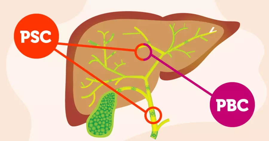
Primary Biliary Cholangitis, commonly called PBC, is a chronic autoimmune disease in which the immune system destroys the small bile ducts within the liver. Bile ducts are tubes that carry bile. Bile is produced by the liver. Bile helps digest fats. Bile also removes bilirubin and cholesterol from the body. When bile ducts are destroyed, bile cannot flow out of the liver. Bile accumulates in the liver. Bilirubin accumulates in blood. Jaundice develops. Cholestasis is the backup of bile. PBC is characterized by progressive destruction of intrahepatic bile ducts. The small bile ducts within the liver are damaged. The medium and large bile ducts are not affected. This distinguishes PBC from Primary Sclerosing Cholangitis (PSC) where large ducts are affected. Primary Biliary Cholangitis affects approximately one to two million people worldwide. The disease predominantly affects women. Women comprise about ninety percent of PBC patients. Men are affected but much less frequently. The disease typically develops in middle-aged to older women. Most common between ages forty and sixty. However, PBC can develop at any age. Juvenile PBC occurs in children but is very rare. Primary Biliary Cholangitis is caused by abnormal immune activation against bile duct epithelial cells. The exact trigger is not known. Genetic predisposition appears important. Environmental factors may trigger disease in susceptible people. Infections may play a role. The immune system produces antibodies against bile duct components. Anti-mitochondrial antibodies develop in about ninety percent of PBC patients. These highly specific antibodies attack mitochondrial antigens in bile duct cells. T cells infiltrate bile ducts. The immune attack causes inflammation. Bile duct cells are damaged and destroyed. Bile ducts narrow. Bile flow is progressively impaired. Cholestasis develops. Bilirubin accumulates. Bilirubin is toxic. Pruritus develops from bilirubin. Portal inflammation spreads. Eventually, cirrhosis can develop. Cirrhosis causes liver failure. Early diagnosis and appropriate treatment are crucial for preventing progression to cirrhosis. Ursodeoxycholic acid treatment slows disease progression. Many people with early PBC achieve good long-term survival. Understanding Primary Biliary Cholangitis helps with early recognition and appropriate management to prevent progressive liver damage and cirrhosis.
How Does Immune Attack on Bile Ducts Cause Primary Biliary Cholangitis?
To understand Primary Biliary Cholangitis, we need to learn about bile ducts and the immune system. The liver produces bile continuously. Bile contains cholesterol, bilirubin, bile salts, and other substances. Bile drains through bile ducts. Small bile ducts within the liver. Medium-sized bile ducts. The common bile duct exits the liver. The bile duct delivers bile to the intestine. Bile is stored in the gallbladder. Bile is released when eating. Bile helps digest fats. In Primary Biliary Cholangitis, the immune system becomes abnormally activated. B cells produce autoimmune antibodies. Anti-mitochondrial antibodies develop. These antibodies are highly specific for PBC. Anti-mitochondrial antibodies attack pyruvate dehydrogenase complex. This enzyme is found in mitochondria of bile duct cells. The antibodies bind to bile duct cells. Complement is activated. Inflammation occurs. Bile duct cell death follows. T cells infiltrate bile ducts. CD8 positive T cells attack bile duct epithelial cells. CD4 positive T cells help coordinate immune response. Inflammatory cytokines are produced. Interleukin-2, interleukin-6, TNF-alpha increase. These inflammatory mediators activate more immune cells. The inflammation spreads. Portal inflammation develops. The portal regions are areas between liver lobules. Portal inflammation is characteristic of PBC. Lymphocytes infiltrate the portal regions. Granulomas form. Granulomas are collections of inflammatory cells. Granulomas are characteristic of PBC. They are present in about ninety percent of PBC cases. The granulomas contain bile duct remnants. Bile ducts are progressively destroyed. The bile duct destruction is progressive. Early PBC shows inflammation and granulomas. Progressive PBC shows progressive bile duct loss. Advanced PBC shows extensive bile duct loss. Bile duct loss leads to ductopenia. Ductopenia is the loss of bile ducts. Cholestasis worsens. Bile flow is progressively impaired. As bile ducts are destroyed, remaining ducts may attempt to compensate. However, with progressive destruction, compensation fails. Cholestasis becomes severe. Bilirubin accumulates progressively. Bilirubin levels increase. Jaundice develops. Pruritus develops from bilirubin and bile acid accumulation. Fibrosis develops as a consequence of chronic cholestasis and inflammation. Bile duct injury triggers fibroblast activation. Collagen accumulates. Portal fibrosis develops. Bridging fibrosis. Eventually, cirrhosis. Cirrhosis is characterized by widespread fibrosis. The liver becomes hardened and scarred. Liver function progressively declines. Portal hypertension develops with cirrhosis. The systemic nature of the immune activation affects other organs. Other autoimmune diseases may coexist with PBC. Sjögren’s syndrome in about fifty percent. Thyroid disease in about twenty percent. Celiac disease. Rheumatoid arthritis. The association with other autoimmune diseases suggests a systemic immune disorder. Understanding the immune mechanisms has led to development of immunosuppressive and cholestatic treatments.
What Are the Main Symptoms and Signs of Primary Biliary Cholangitis?
Primary Biliary Cholangitis causes variable symptoms depending on disease stage and severity. Many people are asymptomatic in early stages. Symptoms develop gradually in most cases. Pruritus, or itching, is the most common symptom. Severe itching. Generalized itching affecting entire body. More pronounced on palms and soles. Worse at night. Sleep disruption from itching. Itching severely impacts quality of life. Pruritus may appear before jaundice. Pruritus occurs from bile acid and bilirubin accumulation in skin. Fatigue is very common. Profound exhaustion. Disproportionate to activity level. Systemic inflammation and cholestasis contribute. Fatigue is often the most disabling symptom. Fatigue limits work capacity and activities. Cognitive changes may accompany fatigue. Difficulty concentrating. Memory problems. Mental slowness. Jaundice develops. Yellowing of skin. Yellowing of sclera. Bilirubin accumulation. Indicates significant cholestasis. Dark urine. Bilirubin in urine. Pale stool. Reduced bile in intestine. Abdominal pain is common. Right upper quadrant pain. Liver pain. Tenderness over liver region. Pain with palpation. The pain may be mild or severe. Abdominal bloating. Abdominal fullness. Gas and bloating. Gastrointestinal discomfort. Nausea may occur. Appetite loss. Reduced food intake. Weight loss from reduced appetite. Joint pain occurs in some. Arthralgia. Non-erosive arthritis. Usually joints not permanently damaged. However, pain significant. Myalgia, or muscle pain, may occur. Muscle aches. Generalized body aches. Fever may occur. Low-grade fever. Indicates active inflammation. Transient fever. Xanthomas develop. Lipid deposits in skin. Yellow nodules on eyelids. Yellow deposits on skin. Occur in advanced disease. From cholestasis and lipid accumulation. Xanthomas may regress with treatment. Portal hypertension signs develop with cirrhosis. Spider angiomas. Small dilated blood vessels. Radiating pattern. Appear on face, chest, hands. Indicate portal hypertension. Palmar erythema. Redness of palms. Vasodilation from liver disease. Ascites develops. Abdominal fluid accumulation. Abdominal swelling. Indicates advanced disease. Edema of legs. Leg swelling. Fluid retention. Hepatomegaly occurs. Liver enlargement. Palpable below rib cage. Tenderness. Splenomegaly occurs. Spleen enlargement. From portal hypertension. Variceal bleeding risk. Esophageal varices. Vomiting blood. Melena, or tarry stool. Life-threatening hemorrhage. Medical emergency. Encephalopathy can develop. Confusion. Mental slowness. Personality changes. Serious symptom. Indicates advanced liver disease. Bone disease signs. Osteoporosis from cholestasis. Bone pain. Fracture risk. Vitamin D deficiency. Hypercalcemia occasionally occurs. From granulomatous disease. Calcium elevation. Hypervitaminosis D from granulomas. Sicca symptoms occur in some. Dry eyes. Dry mouth. From associated Sjögren’s syndrome. Thyroid symptoms may occur. Hypothyroidism. Fatigue. Weight gain. Cold intolerance. From associated thyroid disease. The symptoms vary widely. Some have minimal symptoms for years. Others develop significant symptoms rapidly. Many are asymptomatic initially. Diagnosis often made on screening. Early diagnosis prevents progression.
How is Primary Biliary Cholangitis Detected and Diagnosed?
Primary Biliary Cholangitis is diagnosed through a combination of clinical findings and specific laboratory and imaging tests. Early diagnosis is important for preventing progression. Clinical history is important. Pruritus and fatigue. Jaundice in some. Abdominal discomfort. Progressive symptoms. Risk factors for autoimmune disease. Female gender. Middle-aged. Coexisting autoimmune diseases. Physical examination reveals characteristic findings. Jaundice if present. Hepatomegaly. Splenomegaly. Spider angiomas if portal hypertension. Palmar erythema if liver disease. Xanthomas if advanced disease. Laboratory tests are crucial. Alkaline phosphatase markedly elevated. Markedly increased. Often disproportionately elevated compared to transaminases. Alkaline phosphatase elevation is characteristic. Gamma-glutamyl transferase, or GGT, elevated. Cholestasis markers. Bilirubin elevated. Total and direct bilirubin. Indicates cholestasis. Aspartate aminotransferase, or AST, mildly elevated. Alanine aminotransferase, or ALT, mildly elevated. Usually less elevated than in hepatitis. AST and ALT elevation mild in PBC. This contrasts with autoimmune hepatitis where transaminase elevation is marked. Albumin. Usually preserved initially. Low albumin indicates advanced disease. Liver synthetic dysfunction. Prothrombin time, or PT. INR, or international normalized ratio. Usually preserved initially. Prolongation indicates advanced liver disease. Bilirubin levels. Correlate with disease severity. Progressive elevation indicates disease progression. Platelet count. Low platelets suggest portal hypertension. Anti-mitochondrial antibodies are diagnostic. Present in about ninety percent of PBC. Highly specific for PBC. Presence confirms diagnosis. Titers may not correlate with disease severity. Anti-mitochondrial antibody subtypes. M2 most common. M2 antibodies most specific. Anti-centromere antibodies. Present in some. Associated with limited disease. Better prognosis. Anti-smooth muscle antibodies. May be present. Indicates overlap features. Anti-nuclear antibodies. Some positivity. Rheumatoid Factor. Usually negative. Anti-CCP antibodies. Usually negative. Immunoglobulin levels. IgM often elevated. Elevated IgM supports diagnosis. But not specific. Liver ultrasound. Assesses liver size and echotexture. Looks for cirrhosis. Assesses portal hypertension. Spleen size. Ascites. Doppler ultrasound assesses portal vein patency. Elastography. Transient elastography measures liver stiffness. Indicates degree of fibrosis. Non-invasive assessment of cirrhosis. FibroScan. Acoustic radiation force impulse. Liver biopsy confirms diagnosis. Shows granulomatous inflammation. Portal inflammation. Bile duct destruction. Granulomas in portal regions. Degree of fibrosis assessment. Stages from 0 to 4. Stage 4 is cirrhosis. Biopsy helps assess severity. However, biopsy invasive. Not always necessary if clinical and laboratory features typical. Anti-mitochondrial antibody positivity alone highly suggestive. CT or MRI if cirrhosis suspected. Detailed imaging. Assesses for cirrhosis. Assesses for portal hypertension. Assesses for complications. The combination of clinical presentation with markedly elevated alkaline phosphatase, elevated bilirubin, positive anti-mitochondrial antibodies, and characteristic liver biopsy findings confirms diagnosis. Early diagnosis allows early treatment. Diagnostic delay allows progression of bile duct destruction.
What Health Complications Do People with Primary Biliary Cholangitis Face?
People with Primary Biliary Cholangitis face complications from progressive bile duct destruction and fibrosis. The complications depend on disease progression and disease control. Cholestasis is the primary complication. Bile accumulates in liver. Bilirubin accumulates in blood. Pruritus develops. Severe pruritus affects quality of life. Xanthomas develop. Cholestasis causes lipid accumulation. Jaundice develops. Visible discoloration. Indicates significant cholestasis. Cholestasis causes malabsorption. Fat-soluble vitamins. Vitamin A deficiency. Night blindness. Vitamin D deficiency. Osteoporosis. Vitamin E deficiency. Neurologic symptoms. Vitamin K deficiency. Coagulopathy. Adequate vitamin supplementation essential. Nutritional deficiency. Steatorrhea. Fatty stools. Malabsorption of fat. Deficiency of other nutrients. Progressive liver fibrosis. Bile duct destruction leads to fibrosis. Progressive scarring. Portal fibrosis. Bridging fibrosis. Eventually cirrhosis. Cirrhosis is irreversible. Once cirrhosis develops, cannot be cured. However, disease progression may be halted with treatment. Cirrhosis causes multiple complications. Portal hypertension. Portal vein pressure elevation. Collateral vessel formation. Esophageal varices. Gastric varices. Ascites. Splenomegaly. Portal hypertension causes life-threatening bleeding. Variceal hemorrhage. Esophageal varices rupture. Massive upper gastrointestinal bleeding. Hematemesis. Melena. Hypovolemic shock. Mortality high without urgent treatment. Ascites. Abdominal fluid accumulation. Abdominal swelling. Functional impairment. Mobility difficulty. Increased intra-abdominal pressure. Hepatic encephalopathy. Ammonia accumulation. Toxic effects on brain. Confusion. Memory loss. Personality changes. Sleep disturbance. Asterixis. Flapping tremor. Coma. Serious complication. Hepatorenal syndrome. Renal failure in liver disease. Acute kidney injury. Oliguria. Life-threatening emergency. Spontaneous bacterial peritonitis. Infection of ascitic fluid. Fever. Abdominal pain. Sepsis. High mortality. Hepatic hydrothorax. Fluid in pleural space. Respiratory compromise. Bone disease. Osteoporosis from cholestasis. Vitamin D deficiency. Malabsorption. Bone pain. Fracture risk. Fat fractures from falls. Coagulopathy. Vitamin K deficiency. Impaired clotting factor synthesis. Prolonged PT/INR. Bleeding risk. Easy bruising. Spontaneous bleeding. Hepatocellular carcinoma risk. Cirrhosis increases HCC risk. Requires surveillance. Ultrasound and AFP monitoring. Cholangiocarcinoma risk. Bile duct cancer. Increased risk in PBC. Especially with PSC overlap. Prognosis worse. Associated autoimmune disease complications. Sjögren’s syndrome. Dry eyes. Dry mouth. Thyroid disease. Hypothyroidism. Fatigue. Weight gain. Celiac disease. Malabsorption. Digestive symptoms. Osteoporosis. Increased fracture risk. From cholestasis. From vitamin D deficiency. From associated autoimmune disease. Malnutrition. Fat-soluble vitamin deficiency. Protein malnutrition. Micronutrient deficiency. Weight loss. Muscle wasting. Depression and psychological impact. Chronic progressive disease. Pruritus severely affecting quality of life. Fatigue limiting activities. Uncertainty about disease progression. Depression common. Anxiety about complications. Fear of liver failure. Mental health support important. Without early diagnosis and appropriate treatment, complications are serious and potentially fatal. With early treatment and good disease control, many complications can be prevented or significantly delayed.
What Treatments Help People with Primary Biliary Cholangitis?
Treatment for Primary Biliary Cholangitis focuses on slowing bile duct destruction and managing cholestasis and its complications. Disease-modifying agents slow bile duct destruction. Ursodeoxycholic acid improves outcomes. Ursodiol. Bile acid. FDA approved for PBC. Slows disease progression. Improves cholestasis markers. Reduces bilirubin elevation. Delays need for transplant. Improves long-term survival. Standard first-line therapy. Generally well-tolerated. Continued long-term. Obeticholic acid. FXR agonist. FDA approved for PBC. Activates farnesoid X receptor. Reduces cholestasis. Improves liver enzymes. Used in addition to ursodeoxycholic acid. More effective combination. Requires careful monitoring. Side effects include pruritus worsening initially. Bezlotoxumab. Monoclonal antibody. FDA approved for PBC. Blocks fibrosis pathway. Shows benefit in clinical trials. Used with ursodeoxycholic acid. Improves outcomes. Fibrates. Fenofibrate. Bezafibrate. Some benefit in PBC. Particularly for pruritus. May improve liver enzymes. Used as adjunctive therapy. Immunosuppressive agents. Azathioprine. Some benefit in PBC. Additional immunosuppression. Used in selected cases. Corticosteroids. Limited role in PBC. May help associated features. But not primary treatment. Side effects limit use. Methotrexate. Some benefit reported. Used in selected cases. Rituximab. B cell depletion. Shows promise. Used in refractory cases. TNF inhibitors. Some benefit in refractory disease. Not first-line therapy. Treatment of pruritus. Ursodeoxycholic acid improves pruritus. Obeticholic acid may worsen initially. Bezafibrate helps pruritus. Rifampicin. Antibiotic. Helps pruritus. Opioid antagonists. Naltrexone. Reduces pruritus. Cholestyramine. Bile acid sequestrant. Binds bile acids. Reduces itching. Phototherapy. Light therapy. Helps pruritus in some. Antihistamines. Reduce itching symptomatically. Not disease-modifying. Vitamin supplementation. Fat-soluble vitamin supplementation essential. Vitamin A. Vitamin D. Vitamin E. Vitamin K. Prevents deficiency complications. Calcium supplementation. Vitamin D and calcium. Prevent osteoporosis. Regular monitoring. Bone density assessment. DEXA scans. Osteoporosis monitoring. Osteoporosis treatment. Bisphosphonates. Maintain bone health. Calcium and vitamin D. Weight-bearing exercise. Adequate protein. Support bone health. Portal hypertension management. Beta-blockers reduce portal pressure. Propranolol. Carvedilol. Reduces variceal bleeding risk. Upper endoscopy screening. Screen for esophageal varices. Treat if present. Endoscopic variceal ligation. Prevents bleeding. Therapeutic intervention if bleeding. Ascites management. Diuretics for fluid overload. Spironolactone. Furosemide. Sodium restriction. Fluid restriction. Monitoring for complications. Regular liver ultrasound. Assess for cirrhosis progression. Assess for HCC if cirrhosis. Alpha-fetoprotein monitoring if cirrhosis. Kidney function monitoring. Watch for hepatorenal syndrome. With appropriate early treatment with ursodeoxycholic acid and obeticholic acid, most PBC patients achieve stable disease. Cholestasis markers improve. Disease progression slows. Cirrhosis development delayed or prevented. Transplant-free survival improves.
Living with Primary Biliary Cholangitis
Living with Primary Biliary Cholangitis requires ongoing medical treatment, symptom management, regular monitoring, lifestyle modifications, and psychological adjustment to a chronic progressive liver disease. For people newly diagnosed with PBC, the diagnosis can bring mixed emotions. Relief that the cause of symptoms is identified. Concern about progressive liver disease. Fear about liver failure. However, understanding that effective treatments exist and disease progression can be slowed offers hope. Patient education about PBC, treatment options, disease course, and monitoring helps people understand their condition. Understanding importance of early treatment is crucial. Medication compliance is essential. Taking ursodeoxycholic acid consistently. Compliance is crucial for disease control. Obeticholic acid if prescribed. Taking other medications as prescribed. Missing doses allows disease activity to increase. Regular monitoring. Liver enzyme testing regularly. Bilirubin monitoring. Prothrombin time monitoring. Guides treatment adjustments. Ultrasound monitoring. Assess for cirrhosis progression. Monitor for complications. Vitamin supplementation compliance. Fat-soluble vitamins. Essential for preventing deficiencies. Continued indefinitely. Bone health monitoring. DEXA scans assess bone density. Bisphosphonates if osteoporosis. Calcium and vitamin D supplementation. Pruritus management. Multiple approaches often needed. Ursodeoxycholic acid. Obeticholic acid. Bezafibrate. Rifampicin. Cholestyramine. Naltrexone. Combination therapy often most effective. Skin care. Moisturizers for dry skin. Avoid irritants. Protect skin from trauma. Cool showers. Avoid hot water. Triggers itching. Regular bathing with gentle soap. Work and school adjustments may become necessary. Severe fatigue may limit work. Pruritus severely affects concentration. Some need flexible work schedules. Some need reduced work hours. Severe disease may require disability. Disability support available. School-age children may need accommodations. Fatigue affecting attendance. Pruritus affecting concentration. Frequent medical appointments. Educational accommodations. School counselor support. Nutrition management. Adequate protein. Adequate calories. Adequate vitamins. Especially fat-soluble vitamins. Calcium and vitamin D. Iron if anemia. Vitamin K adequate for clotting function. If malabsorption, nutritional supplementation. Small frequent meals. Avoid fatty foods if steatorrhea. Adequate hydration. No alcohol. Alcohol toxic to liver. Hepatotoxic medications avoided. NSAIDs avoided. Acetaminophen used with caution. Consult physician about all medications. Stress management. Stress reduces quality of life. Meditation and relaxation. Yoga. Regular moderate exercise. Improves overall health. Reduces stress. Supports cardiovascular health. Bone health. Mental health support crucial. Depression common. Anxiety about disease progression. Fear of liver failure. Pruritus severely affects mood. Counseling helps. Antidepressants may be necessary. Support groups. Connect with others with PBC. Share experiences. Coping strategies. Online communities. Social support from family and friends. Family education about disease. Understanding fatigue and limitations. Support with activity modifications. Emotional support crucial. Dating and relationships affected. Fatigue affects intimacy. Pruritus affects appearance. Sexual dysfunction from disease or medications. Communication helps partners understand. Pregnancy possible but requires medical planning. Disease activity may change during pregnancy. Some medications safe, others not. Close medical monitoring essential. Risk of complications during pregnancy. Intrahepatic cholestasis of pregnancy. Liver decompensation. Regular obstetric and hepatology follow-up. Breastfeeding usually safe. With appropriate early treatment with ursodeoxycholic acid and obeticholic acid, vitamin supplementation, symptom management, stress management, adequate nutrition, alcohol avoidance, psychological support, family and social support, and regular medical monitoring, most people with Primary Biliary Cholangitis can slow disease progression and maintain reasonable quality of life despite the chronic progressive nature of this bile duct autoimmune disease.
Frequently Asked Questions About Primary Biliary Cholangitis
FAQ 1: Is Primary Biliary Cholangitis the same as Primary Sclerosing Cholangitis? No, Primary Biliary Cholangitis and Primary Sclerosing Cholangitis are different diseases. PBC affects small bile ducts within the liver. PSC affects medium and large bile ducts outside and within the liver. PBC predominantly affects women. PSC predominantly affects men. PBC has anti-mitochondrial antibodies. PSC typically negative for anti-mitochondrial antibodies. PSC often associated with inflammatory bowel disease. PBC usually not. PBC has better prognosis than PSC. The distinction is important for treatment. They have different treatments and outlooks.
FAQ 2: Can Primary Biliary Cholangitis be cured? Primary Biliary Cholangitis cannot be completely cured. The underlying autoimmune process cannot be reversed. However, disease progression can be significantly slowed. Ursodeoxycholic acid and obeticholic acid slow bile duct destruction. Many achieve stable disease. Disease progression slows. Cirrhosis development delayed or prevented. With appropriate treatment, many never require liver transplant. Quality of life improves with treatment.
FAQ 3: What is the life expectancy for people with Primary Biliary Cholangitis? Life expectancy varies. Early diagnosis improves prognosis. With early treatment, life expectancy approaches normal. Five-year survival approximately ninety-five percent if diagnosed early. Ten-year survival approximately eighty-five percent. Progression to cirrhosis decreases survival. However, with modern treatment, cirrhosis development often prevented. Most live normal or near-normal lifespans. Transplant-free survival achieved in most.
FAQ 4: Can women with PBC become pregnant? Yes, pregnancy is possible in women with PBC. Disease severity may change during pregnancy. Some improve. Some worsen. Close monitoring essential. Some medications safe, others not. Medical planning before pregnancy important. Regular obstetric and hepatology follow-up. Most can have successful pregnancies. However, complications possible. Intrahepatic cholestasis of pregnancy. Liver decompensation. Close monitoring minimizes risks.
FAQ 5: Are there new treatments being developed for Primary Biliary Cholangitis? Yes, ongoing research into improved PBC treatments. Better understanding of fibrosis mechanisms. Antifibrotic agents in development. Selective farnesoid X receptor agonists. Peroxisome proliferator-activated receptor agonists. Bile acid receptor agonists. JAK inhibitors being studied. IL-17 inhibitors showing promise. Gene therapy approaches being researched. Clinical trials of new medications continue. As new treatments develop, outcomes improve. Fibrosis prevention and reversal strategies being researched.
References and Further Reading
For more information about Primary Biliary Cholangitis, you can visit several trusted and authoritative sources providing detailed information for patients and families dealing with this chronic bile duct autoimmune disease. The World Health Organization at WHO.int provides comprehensive information about Primary Biliary Cholangitis and autoimmune liver diseases. The American Association for the Study of Liver Diseases at AASLD.org offers clinical resources and patient education about PBC and liver diseases. The Primary Biliary Cholangitis Organization at PBCOrg.org provides patient education and support specifically for those with PBC. The American College of Gastroenterology at ACG.care provides resources for gastrointestinal and liver diseases including PBC. MedlinePlus, a service of the National Library of Medicine at MedlinePlus.gov, has detailed medical information about Primary Biliary Cholangitis written in language that patients and families can easily understand without specialized medical knowledge. The five main reference links are: 1) WHO.int – Primary Biliary Cholangitis, 2) American Association for the Study of Liver Diseases, 3) Primary Biliary Cholangitis Organization, 4) American College of Gastroenterology, and 5) MedlinePlus – Primary Biliary Cholangitis.
Disclaimer
This article adapts publicly available information from WHO’s Primary Biliary Cholangitis and liver disease information pages. This content is for informational and educational purposes only and does not constitute medical advice. ObserverVoice.com is a news and information platform — not a healthcare provider. If you or someone you know has been diagnosed with Primary Biliary Cholangitis or shows signs of this condition including pruritus, fatigue, jaundice, dark urine, pale stool, abdominal pain, or other symptoms, please consult immediately with qualified healthcare professionals, hepatologists, and liver disease specialists for proper diagnostic evaluation with liver function tests, alkaline phosphatase and bilirubin testing, anti-mitochondrial antibody testing, and liver biopsy if appropriate, and for appropriate early treatment planning with ursodeoxycholic acid and obeticholic acid. Early diagnosis and early aggressive treatment significantly slow disease progression and prevent cirrhosis and liver failure. For more information, visit WHO.int and ObserverVoice.com.
Observer Voice is the one stop site for National, International news, Sports, Editor’s Choice, Art/culture contents, Quotes and much more. We also cover historical contents. Historical contents includes World History, Indian History, and what happened today. The website also covers Entertainment across the India and World.

