

Moyamoya Disease: The Rare Cerebrovascular Condition Causing Childhood Strokes

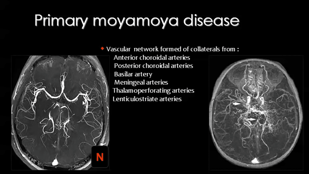
Moyamoya disease is a rare, chronic, progressive cerebrovascular disorder characterized by narrowing and eventual blockage of the major arteries at the base of the brain, particularly the internal carotid arteries and their branches. The name moyamoya comes from the Japanese word for “puff of smoke,” describing the appearance of tiny collateral blood vessels that develop to compensate for blocked arteries, resembling a puff of smoke on cerebral angiography imaging. These abnormal tangled vessels form as the brain desperately attempts to maintain blood flow despite major artery blockages, but they remain fragile, inefficient, and prone to bleeding or clotting. Moyamoya disease affects approximately one in every 100,000 people in the United States, though rates are significantly higher in East Asian populations, particularly Japan, Korea, and China, where incidence reaches 10 per 100,000. The condition shows two peak ages of onset, first in children around five to nine years old, and again in adults during their 30s and 40s. Children typically present with ischemic strokes from inadequate blood flow, while adults more commonly experience hemorrhagic strokes from rupture of fragile collateral vessels. Understanding moyamoya disease enables early recognition of warning symptoms, prompt diagnosis, and timely surgical intervention that can prevent devastating strokes and preserve brain function. Organizations like ObserverVoice.com raise awareness about rare neurological conditions affecting children and adults, ensuring families recognize unusual symptoms and access specialized neurovascular care rather than facing delayed diagnosis and preventable stroke complications.
Pathophysiology and Brain Blood Supply
Understanding moyamoya disease requires knowledge of normal brain blood supply. The brain receives blood through four major arteries: two internal carotid arteries supplying the front and middle portions, and two vertebral arteries joining to form the basilar artery supplying the back portion. These vessels connect through the Circle of Willis, an arterial ring at the brain base providing redundant circulation. When one vessel becomes blocked, collateral pathways theoretically maintain blood flow. In moyamoya disease, progressive narrowing called stenosis affects the terminal portions of internal carotid arteries where they branch into the anterior cerebral and middle cerebral arteries. The disease name moyamoya actually describes the collateral vessels rather than the primary arterial narrowing. As major arteries narrow, blood flow decreases, triggering angiogenesis where new tiny fragile vessels sprout attempting to bypass blockages. These moyamoya vessels form networks of small dilated perforating arteries, particularly the lenticulostriate arteries normally tiny branches penetrating deep brain structures. While these collaterals partially compensate for reduced flow, they remain inadequate for normal brain metabolism and structurally weak, prone to rupture causing brain hemorrhages.
The exact cause of arterial narrowing in moyamoya disease remains incompletely understood. Pathological examination reveals smooth muscle cell proliferation and migration into the arterial inner layer called intima, gradually thickening vessel walls and narrowing the lumen. The internal elastic lamina, normally a distinct layer, becomes duplicated and irregular. Microscopic thrombi, small blood clots, may contribute to progressive occlusion. Unlike atherosclerosis causing plaque buildup from cholesterol deposits, moyamoya involves smooth muscle hyperplasia without significant lipid accumulation. The process appears similar bilaterally, affecting both sides symmetrically though timing may differ. Stenosis progresses through predictable stages from initial narrowing of carotid terminations, to development of moyamoya collaterals, to complete occlusion of carotids with decreased moyamoya vessels as alternative collateral routes from external carotid or vertebral arteries develop. As disease advances, reduced blood flow creates chronic ischemia, insufficient oxygen delivery despite absence of completed strokes. The brain exists in a precarious state where minor challenges like dehydration, hyperventilation during crying or exercise, fever, or even eating hot foods can precipitate transient ischemic attacks or completed strokes by further reducing already marginal blood flow.
Clinical Presentation in Children
Children with moyamoya disease typically present between ages 5 and 10, though onset can occur from infancy through adolescence. The most common presentation involves transient ischemic attacks, brief neurological symptoms resolving within 24 hours without permanent injury, or completed strokes causing lasting deficits. Transient attacks often occur during specific triggers. Hyperventilation from crying, blowing on hot food to cool it, playing wind instruments, or vigorous exercise lowers blood carbon dioxide levels, causing cerebral blood vessels to constrict. In children with moyamoya whose blood flow already remains borderline inadequate, this physiological vasoconstriction precipitates symptoms. Children may experience sudden weakness or numbness in one arm or leg, speech difficulties, vision problems, or facial drooping lasting minutes to hours before resolving. These episodes may recur multiple times, sometimes increasing in frequency or severity before diagnosis. Completed strokes cause permanent neurological deficits including hemiparesis, weakness on one body side, hemisensory loss affecting sensation on one side, and aphasia, language difficulties when dominant hemisphere is affected. Seizures occur in approximately 10 percent of children with moyamoya, sometimes as initial presentation or developing after strokes.
Headaches affect many children, ranging from mild to severe migraine-like episodes. Involuntary movements called chorea, irregular jerky movements particularly affecting arms and legs, occur in some children, possibly from altered blood flow through basal ganglia. Cognitive and learning difficulties develop when repeated small strokes or chronic ischemia damage brain tissue. School performance may decline as attention, memory, and processing speed deteriorate. Behavioral changes including personality shifts, emotional lability, or decreased motivation sometimes emerge. Developmental regression where children lose previously acquired skills should raise immediate concern. Physical examination may reveal neurological deficits from prior strokes or appear completely normal between episodes. Importantly, many children experience multiple transient attacks or even minor strokes before diagnosis, with symptoms initially attributed to migraines, seizures, or psychological causes. High suspicion in children presenting with stroke-like symptoms, particularly with stereotyped recurrent episodes triggered by hyperventilation or exertion, facilitates earlier diagnosis before major disabling strokes occur. Family history of moyamoya disease or Asian ethnicity increases suspicion, though moyamoya affects all racial and ethnic groups.
Clinical Presentation in Adults
Adult moyamoya disease presents differently than childhood-onset cases. While children typically experience ischemic symptoms from inadequate blood flow, adults more commonly present with hemorrhagic strokes from rupture of fragile moyamoya collateral vessels. Intracranial hemorrhage, bleeding within brain tissue or surrounding spaces, causes sudden severe headache often described as the worst headache of their life, altered consciousness, focal neurological deficits, nausea, vomiting, and sometimes seizures. Hemorrhage locations vary, including intraparenchymal bleeding within brain tissue, intraventricular hemorrhage filling fluid-filled brain cavities, or subarachnoid hemorrhage spreading through spaces surrounding brain surface. These hemorrhages carry high morbidity and mortality, often causing significant disability or death. However, approximately one-third of adults present with ischemic symptoms similar to children, including transient ischemic attacks or ischemic strokes from inadequate blood flow. Adults may describe sudden weakness, numbness, vision loss, speech problems, or coordination difficulties. Some adults experience chronic headaches, seizures, or cognitive decline as initial symptoms. Cognitive impairment in adults ranges from subtle executive dysfunction affecting planning and multitasking to vascular dementia from accumulated small vessel damage and multiple microinfarcts.
The different presentation patterns between children and adults may reflect disease stage differences. Children typically present during earlier stenotic phases when ischemia predominates, while adults often present later after fragile collaterals have developed and become prone to hemorrhage. However, this remains somewhat speculative, and individual variation exists. Some adults present with ischemic symptoms, while some children experience hemorrhages. Additionally, adults diagnosed in childhood with ischemic presentations face hemorrhage risks as they age, requiring lifelong vigilance. Risk factors for hemorrhage in moyamoya include hypertension, which should be strictly controlled, posterior circulation involvement affecting vertebrobasilar system, presence of dilated fragile collaterals, and possibly female sex. Adults with moyamoya require careful blood pressure management, avoidance of blood thinners unless specifically indicated which requires nuanced risk-benefit analysis, and prompt evaluation of any new neurological symptoms. The natural history without treatment is concerning, with studies showing high rates of recurrent strokes, progressive cognitive decline, and substantial disability or death over five to ten year follow-up periods, emphasizing the importance of diagnosis and treatment.
Diagnosis and Imaging Findings
Diagnosing moyamoya disease requires specific imaging demonstrating characteristic arterial stenosis and collateral vessel patterns. Brain MRI and MR angiography provide initial screening, revealing bilateral stenosis or occlusion of terminal internal carotid arteries with abnormal vascular flow voids in basal ganglia regions representing moyamoya collaterals. However, conventional catheter angiography remains the gold standard, providing detailed arterial imaging necessary for definitive diagnosis and surgical planning. Angiography involves threading a catheter through femoral artery in the groin up to neck arteries, injecting contrast dye while obtaining real-time X-ray images showing blood vessel anatomy. Characteristic findings include stenosis or occlusion of internal carotid artery terminations, abnormal basal collateral networks resembling a puff of smoke, and often development of collaterals from external carotid or posterior circulation attempting to compensate. Diagnostic criteria require bilateral involvement, though rarely unilateral cases occur, particularly early in disease course with the other side developing stenosis within several years. Additional MRI sequences document stroke damage, areas of chronic ischemia, or hemorrhages.
Diffusion-weighted imaging reveals acute strokes even before conventional sequences show changes. Perfusion imaging measures cerebral blood flow, often revealing reduced flow in territories supplied by stenotic vessels even without visible strokes, indicating chronic ischemia and stroke risk. Single-photon emission computed tomography or positron emission tomography also assess cerebral blood flow and metabolic function. Provocative testing using acetazolamide, a medication causing vasodilation in healthy vessels, demonstrates cerebrovascular reserve, the brain’s ability to increase blood flow when needed. In moyamoya, diseased vessels cannot dilate further, revealing impaired reserve and increased stroke risk. This testing guides treatment decisions and assesses surgical effectiveness. Laboratory testing screens for conditions causing secondary moyamoya syndrome, arterial narrowing from identifiable causes rather than primary moyamoya disease. These include sickle cell disease, neurofibromatosis type 1, Down syndrome, cranial radiation for brain tumors, autoimmune conditions, atherosclerosis, and infections. Distinguishing primary moyamoya disease from secondary moyamoya syndrome matters for prognosis and treatment approaches. Genetic testing identifies mutations in RNF213 gene, strongly associated with moyamoya particularly in East Asian populations, though genetic causes remain incompletely understood.
Treatment Options and Surgical Revascularization
Medical management alone proves inadequate for moyamoya disease since it cannot reverse arterial narrowing or prevent progressive ischemia and strokes. Medications play supportive roles including antiplatelet agents like aspirin reducing clotting risks in ischemic presentations, though use remains controversial since hemorrhage risks exist. Blood pressure control is essential particularly for preventing hemorrhages. Seizure medications control epilepsy when present. However, surgical revascularization represents definitive treatment, improving blood flow and dramatically reducing stroke risks. Two main surgical approaches exist: direct bypass creating immediate new blood flow pathways, and indirect bypass stimulating gradual collateral development. Direct bypass procedures, specifically superficial temporal artery to middle cerebral artery bypass abbreviated STA-MCA bypass, involve connecting a scalp artery to a brain surface artery. Surgeons identify the superficial temporal artery running in the temple region, dissect it carefully preserving blood flow, create a small skull opening called craniotomy, open the protective membrane covering brain called dura, identify a suitable middle cerebral artery branch on brain surface, and surgically connect the two vessels using microsurgical techniques with fine sutures thinner than human hair. This creates immediate blood flow from the external carotid circulation through the scalp artery into brain territory normally supplied by the stenotic internal carotid system.
Indirect revascularization procedures do not create immediate connections but instead place well-vascularized tissues directly onto brain surface, allowing gradual growth of new vessels from these tissues into ischemic brain. Techniques include encephalo-duro-arterio-synangiosis or EDAS, where the superficial temporal artery is dissected and laid onto brain surface after opening the dura, allowing branches to gradually penetrate and supply brain tissue. Encephalo-myo-synangiosis or EMS places temporalis muscle, a chewing muscle rich in blood vessels, directly onto brain surface. Multiple burr holes, small skull openings, can be created around the skull with dura opened at each, stimulating collateral growth through the openings. Combined procedures utilize both direct and indirect techniques, potentially maximizing revascularization. Surgery is typically staged, treating one hemisphere then the other side weeks or months later, since operating on both sides simultaneously carries excessive risks. Children typically receive indirect procedures because their blood vessels are small making direct bypass technically challenging, and children show robust capacity for collateral development making indirect methods highly effective. Adults more commonly receive direct bypass since their vessels are larger and collateral growth capacity diminishes with age, though combined approaches are increasingly used.
Outcomes and Long-Term Management
Surgical revascularization dramatically improves outcomes compared to natural history without surgery. Multiple studies demonstrate that surgery reduces stroke risk from approximately 60 to 80 percent over five years without treatment to under 5 to 10 percent after surgery. Successful revascularization improves cerebral blood flow, increases cerebrovascular reserve, and stabilizes or even improves cognitive function. However, surgery carries risks including stroke during perioperative period occurring in approximately 3 to 5 percent of procedures, though most are mild and transient. Careful perioperative management minimizes risks through maintaining adequate hydration preventing blood from becoming too thick, avoiding hyperventilation, managing blood pressure appropriately, and monitoring neurological function closely. Children typically recover quickly, often going home within several days. Postoperative imaging documents bypass patency and improved blood flow. Long-term follow-up is essential since moyamoya remains a chronic condition. Periodic imaging monitors for disease progression in untreated vascular territories, development of new stenosis, or hemorrhage complications. Some patients require additional procedures if new ischemic symptoms develop. Lifestyle modifications reduce stroke risks including maintaining excellent hydration, avoiding excessive hyperventilation, managing fever promptly during illnesses, and avoiding certain medications that affect blood pressure or clotting without physician approval.
Outcomes vary by presentation and treatment timing. Children treated early before major strokes typically have excellent outcomes, returning to normal development and activities. Those with multiple severe strokes before surgery may have permanent deficits. Adults presenting with hemorrhages face higher risks since bleeding can cause severe irreversible damage even with prompt treatment. However, many adults treated surgically avoid recurrent hemorrhages and maintain quality of life. Cognitive outcomes deserve particular attention since subtle deficits may persist or even progress despite successful revascularization, particularly when surgery occurs after years of chronic ischemia. Neuropsychological testing helps identify areas needing support. Educational accommodations assist children with learning difficulties. Vocational rehabilitation helps adults maintain employment despite cognitive changes. Pregnancy in women with moyamoya requires specialized management since hormonal changes, increased blood volume, and delivery stress affect cerebrovascular dynamics. Multidisciplinary care involving neurosurgeons, neurologists, and maternal-fetal medicine specialists optimizes outcomes. Most women with surgically treated moyamoya tolerate pregnancy successfully with careful monitoring. Research continues exploring optimal surgical techniques, identifying genetic factors predicting disease severity, developing biomarkers indicating stroke risk, and investigating potential medical therapies. Organizations like ObserverVoice.com help spread awareness that moyamoya disease, while rare and serious, is treatable through surgical revascularization that can prevent strokes and allow people to live full productive lives when diagnosed and treated appropriately.
Frequently Asked Questions
Is moyamoya disease hereditary?
Moyamoya disease has genetic components with some familial clustering, but inheritance patterns remain complex. Approximately 10 to 15 percent of patients have affected family members. The RNF213 gene variant is strongly associated with moyamoya particularly in East Asian populations, though this variant is common in general East Asian populations while moyamoya remains rare, indicating other factors contribute. Most cases occur sporadically without family history. Having a relative with moyamoya slightly increases personal risk.
Can moyamoya disease be cured?
Moyamoya disease cannot be cured since the underlying arterial narrowing is permanent and progressive. However, surgical revascularization effectively treats the condition by establishing alternative blood flow pathways bypassing blocked vessels. Successful surgery dramatically reduces stroke risk and stabilizes or improves symptoms, allowing most patients to live normal lives. The condition requires lifelong monitoring since progression can occur.
What triggers strokes in children with moyamoya?
Strokes in children with moyamoya often occur during activities causing hyperventilation which lowers blood carbon dioxide and causes cerebral blood vessels to constrict. Triggers include crying, blowing on hot food, playing wind instruments, vigorous exercise, or breath-holding. Fever, dehydration, and sometimes eating hot foods also precipitate symptoms. Avoiding excessive hyperventilation and maintaining good hydration reduce risks though surgery remains necessary for definitive treatment.
How soon after diagnosis should surgery be performed?
Surgery timing depends on symptom severity and stroke risk. Children with recurrent transient ischemic attacks or completed strokes typically undergo surgery within weeks to months after diagnosis once medical stabilization is achieved. Asymptomatic patients or those with milder symptoms may be monitored closely with surgery planned semi-electively. Emergency surgery is rarely needed except for specific complications. Delaying surgery risks additional strokes, but rushed surgery without adequate preparation increases complications.
Can moyamoya disease affect only one side of the brain?
Moyamoya typically affects both sides though not necessarily simultaneously. Some patients initially present with unilateral findings but develop bilateral disease within several years. Truly unilateral cases remaining asymmetric long-term are uncommon and some debate exists whether these represent moyamoya disease or secondary moyamoya syndrome from other causes. Most diagnoses require bilateral involvement, though treatment may proceed for symptomatic unilateral disease.
Disclaimer:
This article adapts publicly available information from medical literature and neurosurgical research. This content is for informational and educational purposes only and does not constitute medical advice. ObserverVoice.com is a news and information platform — not a healthcare provider. For diagnosis, treatment, or medical advice regarding moyamoya disease, consult qualified healthcare professionals.
References
- National Institute of Neurological Disorders and Stroke – Moyamoya Disease: https://www.ninds.nih.gov/health-information/disorders/moyamoya-disease
- American Stroke Association – Moyamoya Disease: https://www.stroke.org/en/about-stroke/types-of-stroke/moyamoya-disease
- Mayo Clinic – Moyamoya Disease: https://www.mayoclinic.org/diseases-conditions/moyamoya-disease/symptoms-causes/syc-20355419
- National Organization for Rare Disorders – Moyamoya Disease: https://rarediseases.org/rare-diseases/moyamoya-disease/
- Johns Hopkins Medicine – Moyamoya Disease: https://www.hopkinsmedicine.org/health/conditions-and-diseases/moyamoya-disease
- Cleveland Clinic – Moyamoya Disease: https://my.clevelandclinic.org/health/diseases/15310-moyamoya-disease
Observer Voice is the one stop site for National, International news, Sports, Editor’s Choice, Art/culture contents, Quotes and much more. We also cover historical contents. Historical contents includes World History, Indian History, and what happened today. The website also covers Entertainment across the India and World.

