
Glaucoma: The ‘Silent Thief of Sight’ and Why Regular Pressure Checks Save Vision
Vision is something most people take entirely for granted — until it begins to disappear. Glaucoma earns its nickname “the silent thief of sight” precisely because it takes vision gradually, painlessly, and almost always without warning. By the time most people notice something is wrong, a significant and irreversible portion of their sight has already gone.
Glaucoma is the leading cause of irreversible blindness worldwide. The World Health Organization estimates that over 80 million people globally live with the condition, and that number continues to rise. Yet the most alarming fact about glaucoma is not its prevalence — it is how preventable its worst consequences are. Regular eye pressure checks catch glaucoma early, and early treatment saves vision that late detection cannot recover.
What Is Glaucoma?
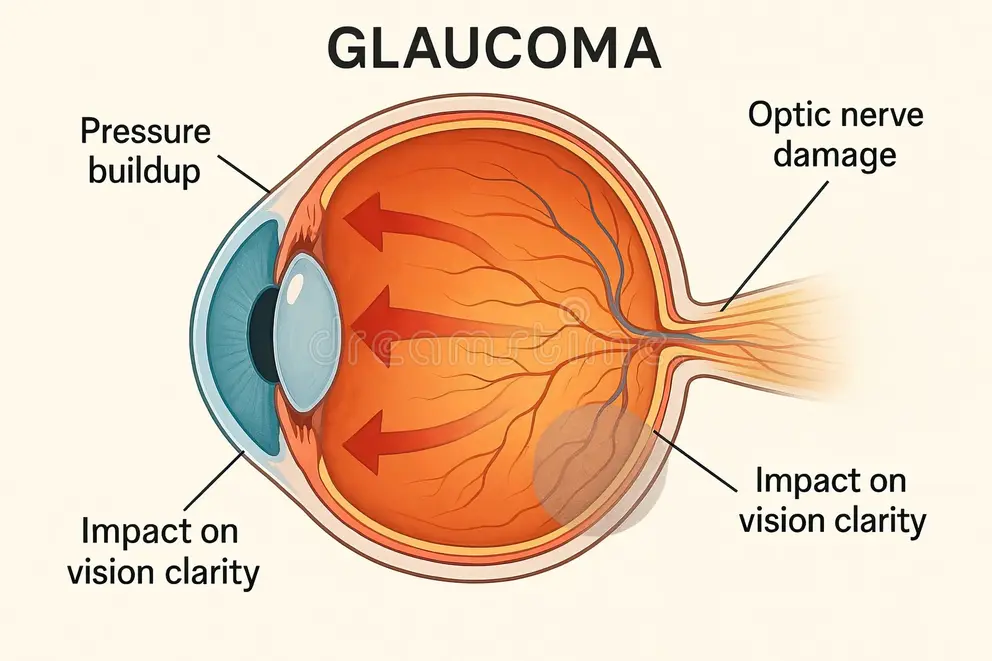
Glaucoma is not a single disease. It is a group of eye conditions that damage the optic nerve — the cable connecting the eye to the brain — typically due to elevated pressure inside the eye. When the optic nerve sustains enough damage, vision loss follows, beginning at the edges and progressing inward.
How the Eye Builds Pressure
The eye constantly produces a clear fluid called aqueous humour. This fluid nourishes internal eye structures and then drains out through a mesh-like channel called the trabecular meshwork, located at the angle where the iris meets the cornea. When this drainage system works properly, fluid pressure inside the eye stays balanced.
When drainage slows or stops, fluid accumulates and pressure builds. Clinicians measure this internal eye pressure in millimetres of mercury, abbreviated as mmHg. Normal intraocular pressure typically falls between 10 and 21 mmHg. Consistently elevated pressure — or even pressure within the normal range in susceptible individuals — can progressively damage the optic nerve.
Why Optic Nerve Damage Is Permanent
The optic nerve contains over one million nerve fibres that carry visual information from the retina to the brain. Unlike many tissues in the body, optic nerve fibres do not regenerate once damaged. Every fibre lost represents permanent, irreversible vision loss.
This irreversibility is why early detection is so critical. Treatment can halt or significantly slow the progression of glaucoma. However, no treatment currently available can restore vision that glaucoma has already destroyed.
Types of Glaucoma
Understanding the different types of glaucoma helps explain why the condition presents so differently across individuals and why screening approaches vary.
Open-Angle Glaucoma
Open-angle glaucoma is the most common form, accounting for approximately 70% to 90% of all glaucoma cases. In this type, the drainage angle between the iris and cornea remains open and appears normal. However, the trabecular meshwork gradually becomes less efficient at draining fluid, causing pressure to rise slowly over months and years.
Open-angle glaucoma develops and progresses almost entirely without symptoms. Vision loss begins in the peripheral — or side — visual field and advances so gradually that the brain compensates, masking the loss until it becomes severe. This symptomless progression explains why so many people with open-angle glaucoma remain undiagnosed for years.
Angle-Closure Glaucoma
Angle-closure glaucoma occurs when the iris bulges forward and physically blocks the drainage angle. This blockage can occur gradually, as in chronic angle-closure glaucoma, or suddenly, producing an acute attack.
An acute angle-closure attack is a medical emergency. It produces sudden, severe eye pain, blurred vision, halos around lights, headache, nausea, and vomiting. Intraocular pressure can spike dramatically within hours. Without immediate treatment, permanent vision loss can occur within days. Anyone experiencing these symptoms must seek emergency care without delay.
Normal-Tension Glaucoma
Normal-tension glaucoma challenges the assumption that elevated eye pressure always explains optic nerve damage. In this form, the optic nerve sustains progressive damage despite intraocular pressure remaining within the statistically normal range.
Researchers believe that increased optic nerve sensitivity to pressure, compromised blood flow to the nerve, or other vascular factors drive normal-tension glaucoma. This type is more common in people of East Asian heritage and in people with a history of low blood pressure or certain cardiovascular conditions.
Secondary and Congenital Glaucoma
Secondary glaucoma develops as a consequence of another eye condition or systemic factor. Eye injuries, steroid medication use, diabetes-related eye disease, and uveitis — inflammation inside the eye — can all elevate intraocular pressure and cause secondary glaucoma.
Congenital glaucoma is present from birth and results from abnormal development of the eye’s drainage system. It is rare but serious, causing symptoms in infants including excessive tearing, light sensitivity, and cloudy corneas. Early surgical intervention produces the best outcomes in congenital glaucoma.
Who Is at Risk of Glaucoma?
Anyone can develop glaucoma, but certain factors significantly increase the risk. Identifying personal risk factors helps people understand when to prioritise eye pressure screening and how frequently to attend follow-up examinations.
Age as a Primary Risk Factor
Age is one of the strongest risk factors for glaucoma. The condition becomes significantly more common after the age of 60. However, glaucoma can develop at any age, including in children and young adults, which is why awareness extends beyond older populations.
After age 40, the risk of glaucoma rises meaningfully with each decade. People over 60 should receive regular glaucoma screening as a standard component of eye healthcare, regardless of current symptom status.
Family History and Genetics
Glaucoma runs in families. People with a first-degree relative — a parent, sibling, or child — diagnosed with glaucoma carry a four to nine times higher risk than those without a family history. This elevated risk reflects shared genetic variants affecting drainage system anatomy, optic nerve resilience, and intraocular pressure regulation.
Knowing family history is clinically valuable. People with affected relatives should begin regular eye pressure checks earlier than the general population and should inform their eye care provider of their family history at every examination.
Ethnic Background and Glaucoma Risk
Research consistently demonstrates differences in glaucoma prevalence and progression across ethnic groups. People of African heritage face a significantly higher risk of developing open-angle glaucoma — approximately three to four times higher than people of European heritage — and tend to develop it at younger ages with more rapid progression.
People of East Asian heritage carry elevated risk of angle-closure glaucoma and normal-tension glaucoma. Hispanic and Latino communities face increasing glaucoma burden as population demographics shift. Culturally sensitive screening programmes that reach all communities at elevated risk are an important public health priority.
Additional Risk Factors
Elevated intraocular pressure is the most significant modifiable risk factor. Even within the statistically normal range, higher pressures increase risk compared to lower pressures. Other contributing factors include thin central corneas — which affect both pressure measurement accuracy and optic nerve susceptibility — high myopia, or short-sightedness, which stretches and thins retinal and optic nerve tissue, and certain cardiovascular conditions affecting blood flow.
Long-term use of corticosteroid medications — including eye drops, oral steroids, and inhaled steroids — raises intraocular pressure in a significant proportion of people. Anyone using long-term steroid therapy in any form should inform their eye care provider so appropriate monitoring can occur.
How Glaucoma Is Detected
Glaucoma detection relies on a combination of tests rather than any single measurement. Understanding what these tests involve helps people engage confidently with screening and follow-up appointments.
Intraocular Pressure Measurement
Measuring intraocular pressure — a process called tonometry — is the most widely known component of glaucoma screening. The gold standard method is Goldmann applanation tonometry, in which a clinician briefly touches the numbed surface of the eye with a small probe to measure the pressure required to flatten a precise area of cornea.
Non-contact tonometry — the familiar “air puff” test — uses a brief burst of air to flatten the cornea and estimate pressure without physical contact. While less precise than Goldmann tonometry, it provides a practical and accessible screening tool in community settings.
Optic Nerve Examination
Measuring eye pressure alone is insufficient for glaucoma diagnosis. The optic nerve must be directly examined to detect damage. Ophthalmoscopy — examination of the back of the eye using specialised lenses and light — allows clinicians to assess the optic nerve’s appearance, including the ratio of its central depression to its overall disc size.
An enlarged optic cup-to-disc ratio can indicate glaucoma-related nerve fibre loss. Photography of the optic nerve provides a baseline image for comparison at future examinations, making change detection more reliable over time.
Visual Field Testing
Glaucoma damages peripheral vision before central vision, making visual field testing an essential diagnostic tool. Automated perimetry — a test in which the person responds to light stimuli appearing at different locations in their visual field — maps the extent and pattern of any vision loss.
Visual field defects characteristic of glaucoma appear in recognisable patterns corresponding to the distribution of optic nerve fibres affected. Repeated visual field tests over time reveal whether the condition progresses and whether treatment is controlling that progression adequately.
Optical Coherence Tomography
Optical coherence tomography, known as OCT, uses light waves to produce detailed cross-sectional images of the retina and optic nerve. OCT can detect thinning of the retinal nerve fibre layer — a direct measure of optic nerve damage — before visual field defects become detectable on standard testing.
This ability to detect structural damage ahead of functional loss makes OCT increasingly valuable in early glaucoma diagnosis and in monitoring treatment response. Many modern glaucoma clinics incorporate OCT as a standard component of both diagnosis and follow-up.
Why Regular Eye Pressure Checks Are Essential
The case for regular eye pressure checks rests on a fundamental clinical reality: glaucoma causes no symptoms until vision loss is already significant, and the vision it takes cannot return. Screening is the only reliable defence.
The Screening Gap
Research consistently reveals that a substantial proportion of people with glaucoma remain undiagnosed. A landmark population study, the Beaver Dam Eye Study, found that approximately half of people with glaucoma in the community did not know they had the condition. Similar findings emerge from studies across diverse populations worldwide.
This diagnostic gap exists because people with no symptoms have no obvious reason to seek eye care. Targeted screening programmes that reach people based on risk factors — age, family history, ethnic background — can dramatically reduce this gap and detect glaucoma before vision loss becomes severe.
Screening Frequency Recommendations
Eye care organisations generally recommend that adults over 40 without known risk factors attend a comprehensive eye examination at least every two years. People with elevated risk factors — including family history, elevated eye pressure, relevant ethnic background, or previous eye injury — should attend annually or as directed by their eye care provider.
The American Academy of Ophthalmology, the Royal College of Ophthalmologists, and the International Glaucoma Association all endorse risk-stratified screening approaches that ensure those at greatest risk receive the most frequent monitoring.
The Role of Community Screening
Many people with elevated glaucoma risk do not attend regular eye examinations. Barriers include cost, access, awareness, and cultural factors. Community-based screening programmes — offered in pharmacies, community centres, and primary care settings — extend the reach of glaucoma detection beyond traditional ophthalmology clinics.
Mobile screening units and teleophthalmology platforms increasingly enable remote assessment of optic nerve images and risk stratification, connecting underserved communities to glaucoma detection services that geography and resources would otherwise restrict.
Treating Glaucoma: Protecting the Vision That Remains
Glaucoma treatment focuses on lowering intraocular pressure to halt or slow optic nerve damage. The specific treatment approach depends on the type and severity of glaucoma, individual patient factors, and treatment response.
Eye Drop Medications
Pressure-lowering eye drops are the most commonly used first-line treatment for open-angle glaucoma. Several classes of drops work through different mechanisms. Prostaglandin analogues — including latanoprost and bimatoprost — increase fluid drainage from the eye and require only once-daily application. They are among the most effective and widely prescribed glaucoma medications.
Beta-blockers such as timolol reduce fluid production within the eye. Carbonic anhydrase inhibitors, alpha agonists, and rho kinase inhibitors offer additional options for people who do not respond adequately to prostaglandins or who cannot use them due to other health conditions.
Adherence to eye drop regimens is critical but frequently challenging. Many people find daily eye drop use difficult to maintain consistently, and reduced adherence directly reduces treatment effectiveness. Clinicians must discuss barriers to adherence openly and help people find practical solutions.
Laser Treatment
Laser treatment offers effective pressure reduction for several glaucoma types. Selective laser trabeculoplasty, known as SLT, targets the trabecular meshwork to improve fluid drainage in open-angle glaucoma. Research from the LiGHT trial — published in The Lancet — demonstrated that SLT as a first-line treatment was as effective as eye drops for most people and may be more cost-effective over the long term.
Laser peripheral iridotomy treats angle-closure glaucoma by creating a tiny opening in the iris to allow fluid to bypass a blocked drainage angle. This procedure prevents acute angle-closure attacks in people identified as at risk through routine examination.
Surgical Treatment
When eye drops and laser treatment fail to control intraocular pressure adequately, surgery becomes necessary. Trabeculectomy — the most established glaucoma surgical procedure — creates a new drainage pathway for aqueous fluid by surgically removing a small section of trabecular meshwork and creating a filtering bleb under the conjunctiva.
Minimally invasive glaucoma surgeries, known collectively as MIGS, represent a rapidly growing area of surgical innovation. Procedures such as trabecular micro-bypass stents, the Hydrus microstent, and canaloplasty offer pressure reduction with fewer risks and faster recovery than traditional trabeculectomy. MIGS is increasingly combined with cataract surgery, allowing simultaneous treatment of both conditions.
Living with Glaucoma
A glaucoma diagnosis requires long-term engagement with eye care rather than a single treatment episode. Understanding what lifelong glaucoma management involves helps people approach the condition with realistic expectations and effective self-management strategies.
Monitoring and Follow-Up
Regular monitoring is the cornerstone of glaucoma management. Eye pressure checks, visual field tests, and optic nerve imaging at intervals determined by disease severity and treatment response allow clinicians to detect progression and adjust treatment before further vision loss occurs.
People with glaucoma must attend follow-up appointments consistently, even when they feel no symptoms and believe their vision is stable. The absence of symptoms does not indicate the absence of progression. Only objective testing can confirm that glaucoma remains under adequate control.
Lifestyle Factors and Glaucoma
Several lifestyle factors influence intraocular pressure and overall eye health. Regular moderate aerobic exercise reduces intraocular pressure in most people and is a valuable complement to medical treatment. Avoiding prolonged head-down positions — such as inverted yoga poses — reduces transient pressure elevations that may stress already-vulnerable optic nerves.
Caffeine consumption transiently raises intraocular pressure, though the clinical significance of this effect in people on treatment remains debated. Maintaining a healthy cardiovascular system supports adequate blood flow to the optic nerve — particularly relevant in normal-tension glaucoma where vascular factors play a significant role.
Emotional Wellbeing and Support
Living with a condition that causes irreversible, progressive vision loss carries a real psychological burden. Anxiety, depression, and grief over vision already lost are common emotional responses that deserve acknowledgment and support alongside physical treatment.
Glaucoma support organisations — including the Glaucoma Research Foundation, Glaucoma UK, and the Glaucoma Foundation — provide peer support, educational resources, and practical guidance for people managing the condition long term. Connecting with these communities reduces isolation and builds the knowledge and confidence that effective self-management requires.
Frequently Asked Questions
Can glaucoma be cured?
Glaucoma currently has no cure. Treatment cannot restore vision that the condition has already destroyed, and it cannot permanently reverse the underlying changes in the drainage system or optic nerve. However, effective treatment can halt or significantly slow progression in most people, protecting the remaining vision for many years. Early detection combined with consistent treatment gives people with glaucoma the best chance of maintaining functional vision throughout their lives.
Does high eye pressure always mean glaucoma?
No. Elevated intraocular pressure — a condition called ocular hypertension — increases the risk of glaucoma but does not automatically mean glaucoma is present. Conversely, some people develop glaucoma despite having eye pressure within the normal statistical range, as in normal-tension glaucoma. Diagnosis requires a combination of pressure measurement, optic nerve examination, and visual field testing rather than pressure measurement alone.
How often should people get their eye pressure checked?
Recommended screening frequency depends on individual risk factors. Adults over 40 with no known risk factors should attend a comprehensive eye examination every two years. People with elevated risk — including family history of glaucoma, elevated eye pressure, relevant ethnic background, high myopia, or steroid medication use — should attend annually or as directed by their eye care provider. People already diagnosed with glaucoma require monitoring at intervals determined by their treating clinician.
Can diet or supplements prevent glaucoma?
No specific diet or supplement has strong evidence for preventing glaucoma. However, general cardiovascular health — supported by a balanced diet, regular physical activity, and avoiding smoking — supports good blood flow to the optic nerve. Some research has explored the role of antioxidants, omega-3 fatty acids, and certain vitamins in optic nerve health, but no supplement currently has sufficient evidence to recommend for glaucoma prevention or treatment.
Is glaucoma hereditary?
Glaucoma has a significant genetic component. People with a first-degree relative diagnosed with glaucoma face a substantially elevated risk of developing the condition themselves. Several genes associated with elevated intraocular pressure, optic nerve anatomy, and drainage system development have been identified. Having a family history of glaucoma is one of the strongest indications for regular screening, beginning earlier than standard population recommendations suggest.
Can children develop glaucoma?
Yes. Childhood glaucoma, while uncommon, includes congenital glaucoma present from birth and juvenile open-angle glaucoma developing in older children and adolescents. Congenital glaucoma produces distinctive symptoms in infants including excessive tearing, light sensitivity, and cloudiness of the cornea. Juvenile glaucoma may be symptomless, as in the adult form. Both require prompt specialist assessment and typically respond to surgical treatment more readily than adult-onset glaucoma.
Conclusion
Glaucoma steals sight quietly, consistently, and permanently. Its silence is both its most dangerous characteristic and its most compelling argument for regular eye pressure checks. The vision glaucoma takes cannot be returned — but the vision it has not yet reached can almost always be protected.
The tools for protection are available, accessible, and effective. Regular intraocular pressure measurement, optic nerve examination, and visual field testing identify glaucoma before it causes significant harm. Pressure-lowering eye drops, laser treatment, and surgery then maintain that protection across a lifetime of management. The entire strategy rests on one foundation: detection before damage becomes irreversible.
Understanding personal risk, attending regular screening, and engaging consistently with treatment are the three actions that transform a glaucoma diagnosis from a sentence to a manageable condition. In the fight against the silent thief of sight, informed, proactive eye care is the most powerful weapon available.
References
- Retinoblastoma is a malignant tumor arising from immature retinal cells characterized by pediatric predominance, genetic basis (RB1 gene), and variable presentations from unilateral to bilateral disease.
- Professional grading starts with comprehensive site analysis. Surveyors map existing elevations and identify problem zones.
- Kris Gopalakrishnan, co-founder of Infosys, views the convergence of sports and technology as a natural progression.
Disclaimer:
This article is for informational purposes only and does not constitute medical advice. Always consult a qualified healthcare professional for diagnosis, treatment, or any eye health concerns.
Observer Voice is the one stop site for National, International news, Sports, Editor’s Choice, Art/culture contents, Quotes and much more. We also cover historical contents. Historical contents includes World History, Indian History, and what happened today. The website also covers Entertainment across the India and World.