

Thyroid Nodules: When to Worry, When to Watch, and What Tests Tell You

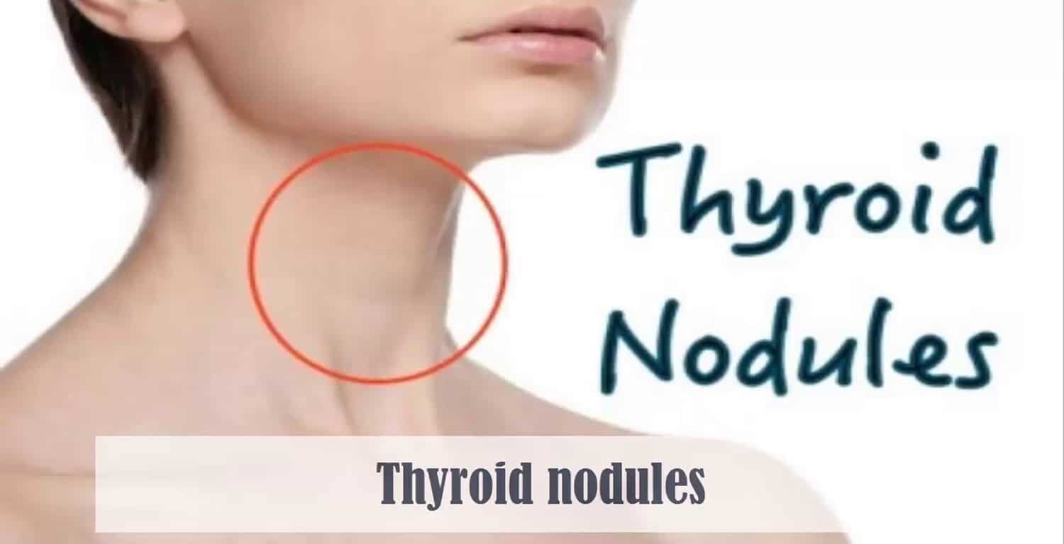
Imagine undergoing a neck ultrasound or CT scan for an unrelated reason. The radiologist reports: “Thyroid nodule identified. Recommend thyroid ultrasound and endocrinology evaluation.” Anxiety floods in. Does this mean cancer? Do I need surgery? Yet thyroid nodules are extremely common—present in approximately 50 percent of ultrasounds. Most are benign. Fewer than 10 percent are malignant. Understanding which nodules require aggressive evaluation versus which can be safely observed prevents both unnecessary surgery and dangerous delays. Proper risk stratification, appropriate testing, and shared decision-making enable optimal management. Thyroid nodules are solid or cystic lesions within the thyroid gland distinct from the surrounding parenchyma. The condition is extremely common—discovered in approximately 50 percent of ultrasounds, 40 percent of CT scans, 16 percent of MRI scans, and approximately 50 percent of autopsy studies. Most thyroid nodules are benign. Fewer than 10 percent are malignant. The challenge is determining which nodules require surgery versus which can be safely observed. Understanding nodule characteristics, risk factors, and appropriate testing enables optimal management decisions. Thyroid nodules affect millions of people. Most are asymptomatic, discovered incidentally. What makes thyroid nodules important is understanding that not all require aggressive intervention. Most benign nodules require only periodic monitoring. Surgery is reserved for nodules with high malignancy risk. Appropriate testing—ultrasound, fine-needle aspiration cytology, molecular testing—guides decision-making. Understanding thyroid nodules prevents both unnecessary surgery and dangerous delays in treating malignancy. In this comprehensive article, we will explore what thyroid nodules are, understand malignancy risk factors, recognize features suggesting benign versus malignant disease, explore diagnostic testing, and discover management strategies for different nodule types.
Understanding Thyroid Nodule Pathophysiology and Malignancy Risk
Before we explore thyroid nodules, we need to understand thyroid physiology and how nodules develop. Thyroid nodule types. Colloid nodules. Most common. Benign. Thyroid follicles. Excessive colloid. Accumulation. Nodule formation. Usually cystic. Or mixed solid-cystic. Non-functional. Thyroid hormone. Not produced. Follicular adenoma. Benign tumor. Encapsulated. Follicular cells. Differentiated. Thyroid hormone. May be produced. Can be hyperfunctioning. Suppressing TSH. Medullary carcinoma. Calcitonin-producing. C cells. Malignant. Genetic. Sometimes. RET proto-oncogene. Mutation. Familial. Multiple endocrine neoplasia. MEN 2. Associated. Papillary carcinoma. Most common thyroid cancer. Approximately 80 percent. Slow-growing. Usually. Excellent prognosis. Often. Lymph node metastasis. Common. Distant metastasis. Rare early. Follicular carcinoma. Second most common. Approximately 10 percent. Encapsulated. Difficult. Distinguish. Benign adenoma. On biopsy. Requires histology. Vascular invasion. Invasion. Follicular carcinoma. Anaplastic carcinoma. Rare. Approximately 1 to 2 percent. Highly aggressive. Rapid growth. Early metastasis. Poor prognosis. Squamous cell. Probably from. Papillary carcinoma. Lymphoma. Thyroid. Rare. Usually B cell. Non-Hodgkin. Metastatic cancer. Other organs. Lung. Kidney. Breast. Others. Metastases. Thyroid. Rare. Benign nodules. Most common. Benign. Colloid nodule. Follicular adenoma. Thyroiditis. Inflammation. Hashimoto’s. Granulomatous. Others. Cysts. Simple. Colloid-filled. Benign. Malignancy risk factors. Age. Younger. Particularly children. Malignancy risk. Higher. Older. Generally. Lower risk. But possible. Gender. Male. Malignancy risk. Higher. Approximately 1.5 to 2 times. Female. Lower risk. Nodule size. Larger nodules. Greater than 4 cm. Malignancy risk. Increases. Smaller. Less than 1 cm. Malignancy risk. Lower. But possible. Growth. Rapid growth. Concerning. Stable. Reassuring. But not completely safe. Family history. Thyroid cancer. Family. RET mutations. Familial medullary carcinoma. MEN 2. Genetic testing. Counseling. Important. Radiation exposure. Head. Neck. Thyroid. History. Malignancy risk. Increased. Childhood. Radiation. Nuclear accident. Atomic bomb. Medical radiation. Prior cancer. History. Malignancy risk. Increased. Lymphocytes. Thyroid infiltration. Thyroiditis. History. Malignancy risk. Slightly increased. Hypothyroidism. Autoimmune. Associated. Slightly increased. Symptoms. Thyroid pain. TSH-suppressive therapy. Levothyroxine. Malignancy risk. Uncertain. Suppression. Sometimes used. Thyroid cancer. History. But not standard. Ultrasound features. Suspicious features. Hypoechogenicity. Darker. Than normal thyroid. Taller than wide. Anterior-posterior. Greater than transverse. Microcalcifications. Small dots. Calcium. Ill-defined margins. Irregular borders. Heterogeneous echotexture. Mixed echogenicity. Exophytic growth. Protruding. Beyond thyroid. Solidity. All solid. Cystic. Liquid-filled. Benign usually. Echogenicity. Isoechoic. Similar. Normal thyroid. Hyperechoic. Lighter. Than normal. Usually benign. Shape. Wider than tall. Benign usually. Taller than wide. Suspicious. Margins. Well-defined. Circumscribed. Benign usually. Ill-defined. Suspicious. Calcification. Coarse. Benign. Micro. Suspicious. The pathophysiology explains nodule formation and malignancy potential.
What Are Thyroid Nodules?
Thyroid nodules are solid or cystic lesions within the thyroid gland. Nodule types. Benign nodules. Most common. Colloid nodules. Benign. Usually cystic. Or mixed. Non-functional. Simple cysts. Fluid-filled. Benign. Pure cysts. Rare. Usually mixed. Follicular adenoma. Benign encapsulated tumor. Non-functional usually. Or hyperfunctioning. Can suppress TSH. Thyroiditis. Inflammatory. Hashimoto’s. Granulomatous. Others. Benign. Nodule-like. Hyperplasia. Benign. Multinodular goiter. Multiple nodules. Benign usually. But cancer. Possible. Any nodule. Malignant nodules. Less common. Papillary carcinoma. Most common thyroid cancer. Approximately 80 percent. Slow-growing. Excellent prognosis. Often. Lymph node metastasis. Common. Distant metastasis. Rare. Follicular carcinoma. Approximately 10 percent. Intermediate growth. Intermediate prognosis. Vascular invasion. Defines. Follicular carcinoma. Anaplastic carcinoma. Rare. Highly aggressive. Rapid growth. Poor prognosis. Medullary carcinoma. Calcitonin-producing. Genetic sometimes. RET proto-oncogene. Mutations. Familial medullary carcinoma. MEN 2. Associated. Lymphoma. Thyroid. Non-Hodgkin. B cell. Rare. Metastatic cancer. Lung. Breast. Kidney. Others. Thyroid. Metastases. Rare. Prevalence. Thyroid nodules. Very common. Approximately 50 percent. Palpation. Physical exam. Approximately 50 percent. Ultrasound. Higher sensitivity. Approximately 40 percent. CT scan. Incidental finding. MRI. Approximately 16 percent. Autopsy. Approximately 50 percent. Most asymptomatic. Discovered incidentally. Malignancy rate. Approximately 5 to 10 percent. Overall. Higher risk features. Malignancy risk increases. Lower risk features. Malignancy risk decreases. Clinical features. Most asymptomatic. No symptoms. Asymptomatic nodules. Most. Incidental findings. Symptoms possible. Neck mass. Palpable. Nodule. Felt. Patient. Physician. Dysphagia. Difficulty swallowing. Large nodule. Compression. Esophagus. Dysphonia. Voice hoarseness. Compression or invasion. Recurrent laryngeal nerve. Stridor. Breathing difficulty. Airway. Compression. Large nodule. Pain. Thyroid. Uncommon. Hemorrhage. Nodule. Cyst. Rupture. Hematoma. Thyroid hormone. Excess. Hyperfunctioning nodule. Rare. Suppresses TSH. Mild hyperthyroidism. Tremor. Palpitations. Heat intolerance. Weight loss. Possible. Complications. Growth. Large nodules. Compression. Esophagus. Trachea. Dysphagia. Stridor. Obstruction. Rare. Hemorrhage. Cystic nodule. Rupture. Hematoma. Sudden enlargement. Pain. Infection. Thyroid cyst. Rare. Abscess. Fever. Pain. Malignant transformation. Benign nodule. Rarely. Becomes malignant. Metastasis. Cancer. Beyond thyroid. Progressive. Thyroid cancer. Untreated. Risks. Compression. Metastasis. Death. Cancer untreated. Long-term. The clinical features influence risk stratification and management decisions.
Recognizing Thyroid Nodules: Clinical Presentation and Risk Assessment
Thyroid nodules have variable presentations guiding clinical management. Asymptomatic discovery. Most common. Incidental finding. Imaging. Ultrasound. CT. MRI. Performed. Other reason. Routine screening. Abnormal. Palpation. Physical. Neck mass. Patient notices. Physician exam. Palpable. Nodule. Enlarging. Family notices. Neck enlargement. Gradual. Progressive. Larger. Over time. Concerning. Rapid growth. Months. Suggests. Malignancy. Possible. Stable. Years. Usually. Benign. But not completely safe. Symptoms from compression. Large nodule. Dysphagia. Difficulty swallowing. Sensation. Mass. Throat. Dysphonia. Voice change. Hoarseness. Stridor. Breathing difficulty. Airway. Obstruction. Hyperthyroidism. Hyperfunctioning nodule. Rare. TSH-suppressed. Tremor. Palpitations. Heat intolerance. Weight loss. Possible. Thyroid pain. Unusual. Nodule. Hemorrhage. Cyst. Rupture. Hematoma. Sudden. Enlargement. Pain. Thyroid. Tenderness. Infection. Abscess. Fever. Pain. Lymph node enlargement. Cervical. Lymphadenopathy. Associated. Papillary carcinoma. Common. Metastatic disease. Distant metastasis. Rare. Lung. Bone. Brain. Other. Advanced. Disease. Constitutional symptoms. Weight loss. Fatigue. Fever. Constitutional. Symptoms. Malignancy. Systemic. Unusual. Local symptoms. Dominant presenting. Cancer risk assessment. Personal risk factors. Age. Younger. Higher. Males. Higher. Radiation exposure. History. Higher. Family history. Thyroid cancer. Genetic. RET mutations. Higher. Prior cancer. Higher. Nodule characteristics. Size. Larger greater than 4 cm. Higher. Growth. Rapid months. Higher. Symptoms. Compression. Hoarseness. Higher. Ultrasound features. Suspicious characteristics. Hypoechogenicity. Microcalcifications. Taller than wide. Ill-defined margins. All solid. Higher. Benign features. Simple cyst. Purely cystic. Isoechoic. Well-defined. Spongiform. Lower. Bethesda classification. Fine-needle aspiration. Results. Non-diagnostic. Atypia of uncertain significance. Follicular lesion. Suspicious. Malignant. Risk. Different. Management decisions. The diverse presentations require systematic risk assessment.
Diagnosis: Comprehensive Nodule Evaluation and Testing
Diagnosing thyroid nodules requires clinical assessment, imaging, and tissue sampling. Clinical history. Nodule discovery. How found. Incidental. Or symptomatic. Growth. Rapid or stable. Symptoms. Compression. Hoarseness. Dysphagia. Pain. Others. Risk factors. Age. Gender. Radiation history. Family history. Prior cancer. Medications. Levothyroxine. TSH suppression. Physical examination. Palpation. Thyroid. Nodule. Location. Size. Consistency. Tenderness. Mobility. Fixed. Suspicious. Lymph nodes. Cervical. Palpable. Metastatic. Possible. Voice. Hoarseness. Recurrent laryngeal nerve. Involvement. Suspicious. Thyroid function tests. TSH. Baseline. Assess function. Low TSH. Hyperfunctioning. Nodule. Possible. Free T4. If TSH abnormal. Thyroid antibodies. TPO. Thyroglobulin. Autoimmune thyroiditis. Associated. Calcitonin. Medullary carcinoma. Suspected. If family history. RET mutations. Genetic counseling. Important. Palpitation. Suspicious features. Ultrasound. First imaging. Sensitivity. High. Approximately 95 percent. Specificity. Moderate. Approximately 50 percent. Thyroid ultrasound. Nodule. Characteristics assessed. Size. Measured. Echogenicity. Hypoechoic. Isoechoic. Hyperechoic. Cystic. Solid. Mixed. Shape. Taller than wide. Wider than tall. Margins. Well-defined. Ill-defined. Irregular. Calcification. Type. Coarse. Micro. Eggshell. Heterogeneity. Homogeneous. Heterogeneous. Vascularity. Doppler. Blood flow. Increased. Suspicious. Exophytic growth. Protruding. Invasion. Surrounding. Structures. Suspicious. Cervical lymph nodes. Size. Shape. Echogenicity. Metastatic. Suspicious. Sonography reporting. TIRADS. Thyroid Imaging Reporting and Data System. Standardizes. Reporting. Risk stratification. TIRADS category. 1. Benign. No FNA. 2. Not Suspicious. No FNA. 3. Mildly Suspicious. FNA. Optional. TSH-dependent. 4. Moderately Suspicious. FNA recommended. 5. Highly Suspicious. FNA recommended. Fine-needle aspiration biopsy. FNA. Ultrasound-guided. Thin needle. 25-27 gauge. Tissue sample. Cells. Smear. Slide. Cytology. Examined. Sensitivity. Approximately 83 to 98 percent. Specificity. Approximately 70 percent. Bethesda classification. Non-diagnostic. Atypia of uncertain significance (AUS). Follicular lesion of undetermined significance (FLUS). Suspicious for follicular neoplasm. Suspicious for malignancy. Malignant. Risk. Escalates. Category. Non-diagnostic. 10 to 40 percent. Malignancy. AUS. 10 to 30 percent. Follicular lesion. 25 to 40 percent. Suspicious malignancy. 50 to 75 percent. Malignant. 95 to 99 percent. Repeat FNA. AUS category. Or follicular lesion. If high suspicion. Surgical recommendation. Genetic testing. Molecular panels. DNA mutations. BRAF. RAS. TP53. PAX8-PPARG. Others. Mutational status. Guides. Malignancy risk. Gene expression classifiers. mRNA expression. Classifiers. Benign. Malignant. Risk stratification. Helpful. Particularly. Follicular. Atypia. Uncertain significance. CT or MRI. Complex cases. Invasion. Surrounding. Structures. Assessment. Distant metastasis. Staging. Cancer. Confirmed. PET-CT. Metastatic disease. Assessment. Staging. Advanced cancer. Diagnostic challenges. Follicular lesion. Follicular neoplasm. FNA. Cannot distinguish. Benign adenoma. Carcinoma. Histology. Required. Vascular invasion. Defines. Carcinoma. Diagnosis. Cannot made. FNA. Core biopsy. Sometimes. Helps. Determines. Invasion. Not always. Definitive. Molecular testing. May help. Mutation status. Malignancy risk. VUS variants. Variants of uncertain significance. Molecular testing. Interpretation. Uncertain. Emerging. Improving. The diagnosis requires systematic clinical assessment and appropriate testing.
Management: Risk-Stratified Approach to Nodule Care
Thyroid nodule management depends on malignancy risk and patient preferences. Benign nodules—low risk. Observation. No treatment. Nodules. Benign features. FNA results. Benign. Malignancy risk. Less than 1 percent. Observation. Recommended. Ultrasound surveillance. Baseline. Initial FNA. Benign result. 6 to 12 months. Then. If stable. 12 to 24 months. Later. If unchanged. Every 2 to 5 years. Indefinite. Or stop. Shared decision-making. Important. Patient preference. Anxiety. Engagement. Consideration. Surgery. Rarely. Benign nodule. Compressive symptoms. Dysphagia. Stridor. Concern. Cosmetic. Psychological. Surgery. Discussion. Risk-benefit. Consideration. FNA repeat. AUS or follicular lesion. Molecular testing. Consider. Gene expression classifier. Mutation analysis. Helps. Malignancy risk. Higher. Molecular testing. Suspicious or malignant. Results. FNA indeterminate. With molecular. Suspicious result. Surgery. Recommended. Benign molecular. Result. Observation. Alternative. Reassurance. Avoid unnecessary. Surgery. Observation advantages. Avoids surgery. Complications. Hypothyroidism. Recurrent laryngeal nerve. Injury. Cost. No treatment. Surveillance. Periodic. Reassurance. Likely benign. Observation drawbacks. Anxiety. Uncertainty. Cancer risk. Small. But present. Inconvenience. Periodic ultrasound. Ongoing. Patient cost. Ultrasound. FNA. Repeat. May be. Multiple times. Shared decision-making. Important. Some patients. Prefer surgery. Peace of mind. Anxiety relief. Others. Prefer observation. Avoid surgery. Avoid treatment. Till necessary. Suspicious or malignant nodules—high risk. Surgery. Recommended. FNA. Suspicious or malignant. Malignancy risk. High. Approximately 50 to 99 percent. Surgery. Thyroidectomy. Partial or total. Recommended. Extent. Depends. Cancer suspicion. Risk. Molecular findings. Papillary. Usually total or near-total. Lobectomy. Possible. If low-risk papillary. Follicular. Total. Vascular invasion. Confirmed or suspected. Anaplastic. Total. Aggressive. Metastatic. Extensive. Medullary. Total. Lymph node dissection. If involved. Radioactive iodine. Post-operative. If high-risk papillary. Metastatic disease. Confirmed or suspected. Surveillance. Post-operative. Thyroglobulin. Marker. Monitoring. Ultrasound. Periodic. Radioactive iodine scan. If indicated. TSH suppression. Levothyroxine. Dose. Often increased. Post-operative. Suppress TSH. Cancer recurrence. Risk. Molecular testing—atypia or follicular. Molecular panel. Gene expression. Helps decision. Higher risk. Genetic mutations. Gene expression. Malignant classifier. Surgery. Recommended. Lower risk. Benign classifier. Observation. Alternative. Repeat FNA. Observation. Multiple options. Shared decision. Monitoring benign nodules. Growth. Small nodule. Less than 1 cm. No FNA. Observation. Ultrasound. Baseline. Then every 6 to 12 months. Initially. If stable. Longer intervals. Eventually. Stability. Reassuring. Growth. Progression. Concerning. Repeat FNA. Growth. Greater than 50 percent. Volume. Or greater than 20 percent. Any dimension. Suggested. Repeat FNA. Some. Recommend. Even stable. Repeat FNA. Validation. Benign results. Continuing. Observation. Fine-needle aspiration—repeat. AUS or follicular. Category. Repeat FNA. 3 to 6 months. Suggested. Concordance. Same result. Observation. Likely. Different. Surgery. Or molecular testing. Molecular panel. Consider. Helps. Risk stratification. Cystic nodules. Pure cysts. Benign. No FNA. Observation. Follow-up ultrasound. Optional. Mixed cysts. Solid component greater than 20 percent. FNA. Suggested. Risk. Increased. Solid component. Biopsy. Sampling. Adequate tissue. Critical. Sonographer. Skill. Important. Nodule. Vascular. Difficult access. Core biopsy. Consider. Surgery. Complications. Hypothyroidism. Most common. Levothyroxine. Replacement. Recurrent laryngeal nerve. Injury. Hoarseness. Voice change. Permanent. Usually temporary. Reinnervation. Weeks to months. Superior laryngeal nerve. Injury. High voice. High notes. Difficult. Permanent sometimes. Hypoparathyroidism. Permanent sometimes. Calcium supplementation. Vitamin D. Necessary. Bleeding. Hematoma. Rare. Infection. Rare. Anesthesia. Risks. Standard surgical. Thyroid cancer outcomes. Papillary. Excellent. 10-year survival. Greater than 95 percent. Often. Localized disease. Follicular. Intermediate. 10-year survival. Approximately 85 percent. Anaplastic. Poor. 10-year survival. Less than 20 percent. Often. Advanced disease. Medullary. Intermediate. Depends. Extent. Genetics. Genetic counseling. Important. RET mutations. Genetic testing. Family. Prophylactic thyroidectomy. Children. RET-positive. MEN 2. Preventive. Surgery. Recommended. The risk-stratified approach prevents both unnecessary surgery and dangerous delays.
Frequently Asked Questions (FAQs)
Q1: Does every thyroid nodule need to be biopsied?
No. Not all nodules require FNA. Benign features. No biopsy necessary. Observation. Appropriate. TIRADS 1 and 2. No FNA. TIRADS 3. Optional. TSH-dependent. TIRADS 4 and 5. FNA recommended. Size matters. Nodules less than 1 cm. Generally. No FNA. Unless suspicious features. Very concerning history. Observation approach. Standard. Most nodules. Safe.
Q2: If my nodule is benign, will I need lifelong monitoring?
Possibly. Benign nodule. Observation. Periodic ultrasound. Years. If stable. Intervals. Lengthen. Eventually. No follow-up. Necessary. Patient preference. Varies. Some reassured. Stop monitoring. Others. Continue. Indefinite. Shared decision-making. Important. Anxiety. Peace of mind. Versus. Inconvenience. Cost. Ongoing surveillance.
Q3: What percentage of thyroid nodules are cancer?
Approximately 5 to 10 percent. Malignant. Approximately 90 to 95 percent. Benign. So most. Benign. Cancer. Possible. Risk. Elevated. Suspicious features. Ultrasound. FNA. Molecular testing. Help. Risk stratification. Better understanding. Malignancy risk.
Q4: Can a benign nodule become cancerous?
Rarely. Benign nodule. Transformation. Malignant. Uncommon. Most cancer. Present initially. Benign appearance. Can sometimes. Low-risk papillary. Remain stable. Years. Never develop. Metastasis. Cancer growth. Rare. Initial benign. Later malignant. Possible. Not standard. Monitoring. Appropriate. Changes. Detected early.
Q5: Do I need surgery if my FNA is uncertain?
Depends. FNA category. AUS or follicular. Surgery. Not always necessary. Observation. Reasonable. Molecular testing. Often. Helpful. Gene expression. Mutation analysis. Guides decision. If molecular. Suspicious. Surgery. Recommended. If benign. Observation. Alternative. Shared decision-making. Important. Individual. Risk tolerance. Patient preference.
Key Takeaways
Thyroid nodules are common. Approximately 50 percent. Ultrasound. Approximately 40 percent. CT. Most benign. Approximately 90 to 95 percent. Cancer risk. Approximately 5 to 10 percent. Benign nodules. Colloid. Most common. Follicular adenoma. Cysts. Simple. Thyroiditis. Malignant nodules. Papillary. Most common cancer. Approximately 80 percent. Follicular. Approximately 10 percent. Anaplastic. Rare. Aggressive. Medullary. Lymphoma. Rare. Malignancy risk factors. Age. Younger. Gender. Male. Radiation. History. Family history. Thyroid cancer. Nodule characteristics. Size. Growth. Ultrasound features. Suspicious. Risk assessment. Clinical history. Nodule features. Ultrasound findings. FNA results. Bethesda classification. Molecular testing. Helps. Risk stratification. Diagnosis. TSH. Thyroid antibodies. Ultrasound. TIRADS classification. FNA. Indicated. TIRADS 4 and 5. Optional. TIRADS 3. Not indicated. TIRADS 1 and 2. Bethesda classification. Non-diagnostic. AUS. Follicular lesion. Suspicious. Malignant. Malignancy risk. Increases. Category. Molecular testing. Gene expression. Mutation analysis. Helps. Indeterminate. FNA results. Management. Benign nodules. Observation. Periodic ultrasound. Surveillance. No treatment. Surgery. Rare. Unless symptoms. Compression. Psychological. Uncertain nodules. Molecular testing. Consider. FNA repeat. Possible. Surgery. Decision. Risk-benefit. Shared. Suspicious or malignant. Surgery. Recommended. Thyroidectomy. Extent. Depends. Cancer. Suspicion. Type. Post-operative. Surveillance. Thyroglobulin. Ultrasound. Radioactive iodine. If indicated. TSH suppression. Cancer. Risk reduction. Outcomes. Papillary carcinoma. Excellent. Greater than 95 percent. 10-year survival. Often. Follicular. Intermediate. Approximately 85 percent. Anaplastic. Poor. Less than 20 percent. Medullary. Intermediate. Genetic counseling. Important. RET mutations. MEN 2. Prophylactic surgery. Children. Recommended. Thyroid nodules—very common—usually benign—management risk-stratified—prevent unnecessary surgery and dangerous delays.
References
- World Health Organization (WHO). “Thyroid Nodule Evaluation and Management.” Retrieved from https://www.who.int/
- American Thyroid Association. “ATA Thyroid Nodule Guidelines.” Retrieved from https://www.thyroid.org/
- American Association of Clinical Endocrinologists. “AACE Thyroid Nodule Guidelines.” Retrieved from https://www.aace.com/
- Radiological Society of North America. “TIRADS Classification System.” Retrieved from https://www.rsna.org/
- Mayo Clinic. “Thyroid Nodules: Diagnosis and Management.” Retrieved from https://www.mayoclinic.org/
- Cleveland Clinic. “Thyroid Nodules: Complete Information.” Retrieved from https://my.clevelandclinic.org/
Related Articles on ObserverVoice.com
Explore more health and science topics on our platform:
- Thyroid Function: Understanding Nodule Development
- Thyroid Cancer: Understanding Different Types and Prognosis
- Ultrasound Imaging: Understanding Thyroid Evaluation
- Fine-Needle Aspiration: Understanding Thyroid Biopsy
- Risk Stratification: Understanding Cancer Risk Assessment
- Thyroid Surgery: Understanding Thyroidectomy and Complications
Disclaimer
This article provides educational information adapted from publicly available health sources including WHO materials. This content is for informational and educational purposes only and does not constitute medical advice, diagnosis, or treatment recommendations. [ObserverVoice.com] is a news and information platform—not a healthcare provider. If you have a thyroid nodule discovered on imaging or physical examination, consult qualified endocrinologists or thyroid specialists for evaluation. Risk stratification using clinical history, ultrasound findings, and appropriate testing (FNA, molecular testing) guides management decisions. Most thyroid nodules are benign and require only periodic monitoring. Not all require surgery. Shared decision-making between patient and physician optimizes management. Risk-based approach prevents both unnecessary surgery and dangerous delays in treating malignancy. Regular surveillance of benign nodules is safe and appropriate. If malignancy suspected, prompt evaluation and treatment recommended. Always seek guidance from licensed healthcare specialists for nodule evaluation and management.
Observer Voice is the one stop site for National, International news, Sports, Editor’s Choice, Art/culture contents, Quotes and much more. We also cover historical contents. Historical contents includes World History, Indian History, and what happened today. The website also covers Entertainment across the India and World.
Follow Us on Twitter, Instagram, Facebook, & LinkedIn

