

Radiation: Understanding Invisible Energy’s Health Effects
Why Medical Imaging Benefits Outweigh Risks When Used Appropriately

Thirty-five-year-old Sarah from Boston sits anxiously in the radiology department waiting room, clutching the referral form from her doctor requesting a CT scan of her abdomen. Her persistent stomach pain needs investigation, but Sarah has read alarming internet articles about radiation from medical scans causing cancer. “Is this test safe?” she asks the radiologic technologist nervously. “Will the radiation harm me? I’ve already had three X-rays this year—am I getting too much radiation?”
Sarah’s concerns reflect widespread confusion about radiation—what it is, how it affects health, when medical radiation is beneficial versus harmful, and how risks compare to benefits. Many people fear all radiation exposure, not realizing we’re constantly exposed to natural background radiation from space, rocks, soil, and even our own bodies. Others underestimate radiation risks, seeking unnecessary medical imaging or not protecting themselves from excessive exposure. Understanding radiation—its sources, health effects, and appropriate use—enables informed decisions balancing legitimate benefits against real but often overstated risks.
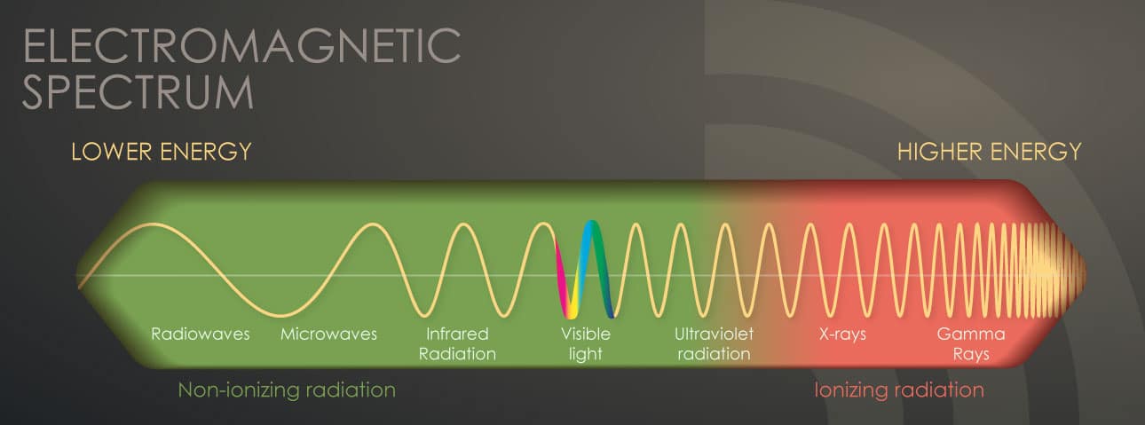
According to the World Health Organization, radiation is energy traveling through space or matter that can be classified as either ionizing (has enough energy to remove electrons from atoms, potentially damaging DNA and causing cancer) or non-ionizing (lower energy that doesn’t remove electrons, generally less harmful). Ionizing radiation sources include natural background radiation (cosmic rays, radon gas, radioactive elements in soil/rocks/food), medical procedures (X-rays, CT scans, nuclear medicine), and man-made sources (nuclear power, occupational exposure, fallout from weapons testing). WHO emphasizes that while high radiation doses clearly cause health problems including cancer, most people’s radiation exposure comes from natural sources and low-dose medical procedures where benefits typically far outweigh small risks when imaging is medically justified.
Understanding Radiation Types
Radiation comes in two fundamental categories with different health implications. Non-ionizing radiation has relatively low energy insufficient to remove electrons from atoms. Types include radiofrequency radiation (radio waves, microwaves, WiFi, cell phones), visible light, infrared radiation (heat), and ultraviolet (UV) radiation from the sun. Most non-ionizing radiation poses minimal health risks at typical exposure levels, though UV radiation causes sunburn and skin cancer with excessive exposure, and very high intensity microwave or radiofrequency radiation can cause tissue heating.
Ionizing radiation possesses enough energy to remove electrons from atoms, creating ions that can damage DNA and other cellular components. This category includes X-rays used in medical imaging, gamma rays from radioactive materials, alpha particles released by radioactive elements (stopped by skin or paper but dangerous if inhaled/ingested), beta particles (penetrate skin but stopped by clothing or thin metal), and neutrons from nuclear reactions. Ionizing radiation’s ability to damage DNA creates both its medical utility (killing cancer cells in radiation therapy) and its health risks (potentially causing cancer when damaging normal cells’ DNA).
Like understanding differences between beneficial nutrition and harmful obesity, distinguishing radiation types and doses helps assess actual risks versus perceived dangers.
Natural and Medical Radiation Exposure
Everyone receives radiation exposure constantly from natural sources. Cosmic radiation from space penetrates Earth’s atmosphere—exposure increases with altitude (airplane passengers and crew receive higher doses, mountain residents more than sea-level dwellers). Terrestrial radiation comes from naturally radioactive elements in rocks, soil, and building materials—levels vary by geography with some areas having naturally high background radiation. Internal radiation occurs because our bodies contain naturally radioactive elements including potassium-40 in bananas and other foods, carbon-14 produced by cosmic rays, and radon gas (and its decay products) inhaled from soil and building materials—radon represents the largest natural radiation source for most people.
Average natural background radiation exposure worldwide is approximately 2-3 millisieverts (mSv) per year, though varies considerably by location—some areas have natural background radiation 10-20 times higher without apparent health effects on residents. Medical radiation has become the largest source of human-made radiation exposure for most people in developed countries. Common medical procedures deliver varying doses: chest X-ray approximately 0.1 mSv (equivalent to 10 days of natural background radiation), mammogram approximately 0.4 mSv, dental X-ray approximately 0.005 mSv, abdominal X-ray approximately 0.7 mSv, CT scan of chest approximately 7 mSv (equivalent to 2-3 years of natural background), CT scan of abdomen approximately 10 mSv, and nuclear medicine scans varying from 1-20 mSv depending on procedure.
For perspective, a cross-country round-trip flight exposes passengers to approximately 0.05 mSv from increased cosmic radiation at altitude. Like occupational health hazards, radiation risks depend on dose, duration, and protective measures.
Health Effects of Radiation
Radiation health effects depend critically on dose and exposure pattern. High-dose acute exposure (received over short time) causes acute radiation syndrome with symptoms including nausea, vomiting, diarrhea, skin burns, hair loss, immune system damage, and potentially death—seen in atomic bomb survivors, Chernobyl accident responders, and rare accidents. Doses above 1,000 mSv (1 sievert) received acutely cause measurable health effects, while doses above 5,000 mSv are usually fatal without intensive medical treatment.
Low-dose chronic exposure (small amounts over extended time) presents more complex risks. Evidence shows radiation can cause cancer years or decades after exposure by damaging DNA in ways that occasionally lead to uncontrolled cell growth. Cancer risk increases with cumulative radiation dose—more radiation means higher cancer risk. However, at low doses (below 100 mSv), cancer risk is very small and difficult to measure statistically. For perspective, a single CT scan (approximately 10 mSv) increases lifetime cancer risk by approximately 1 in 2,000—small compared to the roughly 1 in 3 baseline cancer risk everyone faces.
Children are more sensitive to radiation than adults because their cells divide more rapidly and they have more years to develop radiation-induced cancers. This makes minimizing radiation exposure particularly important for pediatric patients. Pregnant women require special consideration since fetal exposure can potentially cause developmental problems, though risks are primarily concerning at doses much higher than typical medical imaging.
Importantly, radiation therapy for cancer intentionally delivers very high doses (20,000-80,000 mSv) to tumors to kill cancer cells. These doses would be extremely dangerous to the whole body but are carefully targeted to cancerous tissue, with benefits of treating cancer far outweighing radiation risks. Like palliative care balancing treatment and quality of life, medical radiation use requires weighing benefits against risks.
Medical Imaging: Benefits and Risks
Medical imaging using ionizing radiation provides enormous benefits when appropriately used. X-rays and CT scans diagnose fractures, tumors, infections, internal injuries, and countless other conditions enabling appropriate treatment. Nuclear medicine procedures assess organ function, detect cancer spread, and guide treatment decisions. Radiation therapy cures many cancers or provides symptom relief.
However, medical radiation also carries small cancer risks, making appropriate use essential. Justification means ensuring each imaging procedure is medically necessary—that expected diagnostic information will influence patient management. Optimization means using lowest radiation dose that provides adequate diagnostic information through modern equipment with dose-reduction features, appropriate imaging protocols based on patient size and clinical question, and limiting scan coverage to necessary areas.
Alternative imaging when appropriate includes ultrasound (uses sound waves, no ionizing radiation) for many abdominal, pelvic, and vascular studies, MRI (uses magnetic fields, no ionizing radiation) for brain, spine, joint, and soft tissue imaging, and clinical examination sometimes providing sufficient information without imaging.
Unnecessary imaging creates risks without benefits through repeated imaging when one study suffices, imaging unlikely to change management, imaging for reassurance rather than medical indication, and screening tests without evidence of benefit. Like patient safety initiatives minimizing medical harm, radiation protection requires systematic approaches.
Radiation Protection Principles
Radiation protection follows three fundamental principles. Justification means any decision introducing radiation exposure should do more good than harm—medical imaging should provide diagnostic information benefiting patient care. Optimization means radiation doses should be kept as low as reasonably achievable (ALARA principle) while still accomplishing the medical purpose. This involves using appropriate equipment settings, protocols, and techniques minimizing dose without compromising diagnostic quality.
Dose limits apply to occupational exposure (workers in nuclear facilities, radiology departments, research facilities) and public exposure from nuclear facilities or other man-made sources. Medical exposure to patients doesn’t have dose limits because restricting medically necessary imaging would harm patients, but justification and optimization principles apply. Time, distance, and shielding provide practical protection—limiting time near radiation sources, maintaining distance (radiation intensity decreases rapidly with distance), and using shielding (lead aprons in X-ray rooms, concrete walls around nuclear facilities).
For medical patients, radiation protection involves asking providers if imaging is necessary and if alternative non-radiation methods exist, informing providers about recent imaging to avoid unnecessary duplication, ensuring technologists use appropriate child-sized protocols for pediatric patients, and keeping personal records of radiation procedures enabling informed decisions about future imaging.
Sarah’s CT Scan Decision
Sarah’s radiologist explained that her persistent abdominal pain, weight loss, and abnormal blood tests created genuine concern requiring investigation. A CT scan could identify serious conditions including appendicitis, kidney stones, tumors, or inflammatory diseases that other tests cannot detect. “The radiation dose from this CT scan—about 10 millisieverts—increases your lifetime cancer risk by roughly 1 in 2,000,” the radiologist explained. “That’s a small risk compared to the immediate need to diagnose what’s causing your symptoms. If we find something serious that we can treat, the benefit far outweighs the radiation risk. However, if your symptoms were mild and we weren’t worried about serious disease, I’d recommend starting with ultrasound instead.”
Understanding this explanation, Sarah proceeded with the CT scan, which identified appendicitis requiring surgery. “I’m glad I had the scan,” Sarah reflected afterward. “It found the problem and enabled timely treatment. I understand radiation carries small risks, but those risks are tiny compared to benefits when imaging is medically necessary. I also learned to question whether imaging is needed rather than assuming every test is necessary—protecting myself from unnecessary radiation while accepting appropriate medical imaging.”
Dr. Chen, the radiologist, emphasizes broader lessons: “Radiation often triggers irrational fear or cavalier dismissal—both problematic. Yes, radiation can cause cancer at high doses. Yes, we should minimize unnecessary exposure particularly in children. But radiation also enables diagnosing and treating countless medical conditions, saving millions of lives through medical imaging and radiation therapy. The solution isn’t avoiding all radiation—that’s impossible given natural background exposure and would deprive patients of life-saving diagnoses. Rather, we need informed decision-making using radiation when benefits outweigh risks, optimizing doses to minimum levels providing diagnostic information, avoiding unnecessary imaging that provides no medical benefit, and educating patients, providers, and the public about realistic radiation risks and benefits. When we achieve this balance, radiation remains one of medicine’s most powerful tools while minimizing its small but real risks.”
Frequently Asked Questions (FAQs)
Ionizing radiation is energy with sufficient power to remove electrons from atoms, potentially damaging DNA and other cellular components. Types include X-rays, gamma rays, and particles from radioactive materials. Health effects depend on dose: High doses (>1,000 mSv acutely) cause acute radiation syndrome—nausea, vomiting, immune damage, potentially death. Lower doses increase cancer risk years/decades later by damaging DNA occasionally leading to uncontrolled cell growth. Cancer risk increases with cumulative dose, but at low doses (<100 mSv), risk is very small—a 10 mSv CT scan increases lifetime cancer risk by approximately 1 in 2,000 compared to baseline 1 in 3 cancer risk everyone faces. Medical benefits typically far outweigh small risks when imaging is medically justified.
Everyone constantly receives natural background radiation from: cosmic radiation from space (increases with altitude—more during flights, at high elevations), terrestrial radiation from radioactive elements in rocks/soil/buildings (varies by geography), internal radiation from naturally radioactive elements our bodies contain (potassium-40, carbon-14), and radon gas from soil/building materials (largest natural source for most people). Average worldwide natural background is approximately 2-3 mSv annually, though varies considerably by location—some areas have 10-20 times higher natural radiation without apparent health problems. A cross-country flight adds approximately 0.05 mSv from increased cosmic radiation. We can’t avoid natural radiation, and human evolution occurred with this constant exposure.
Medical imaging using ionizing radiation carries small cancer risks but provides enormous benefits when appropriately used. A chest X-ray (0.1 mSv) equals approximately 10 days of natural background radiation. A CT scan (7-10 mSv) equals approximately 2-3 years of natural background. These doses increase lifetime cancer risk slightly—a 10 mSv CT scan by approximately 1 in 2,000. However, when imaging is medically necessary for diagnosing conditions requiring treatment, benefits far outweigh small risks. Problems arise from unnecessary imaging providing no medical benefit while adding radiation exposure. The key is justification—ensuring each imaging procedure is medically necessary—and optimization—using minimum dose providing diagnostic information. Children require special care due to higher radiation sensitivity.
Protect against unnecessary radiation by: (1) Ask providers if imaging is necessary and if non-radiation alternatives (ultrasound, MRI, clinical examination) could provide needed information; (2) Inform providers about recent imaging avoiding unnecessary duplication; (3) Keep personal records of radiation procedures enabling informed decisions about future imaging; (4) For children, ensure technologists use appropriate child-sized protocols minimizing dose; (5) Accept medically justified imaging—avoiding necessary diagnostic tests due to radiation fear can result in missing serious conditions requiring treatment; (6) Limit radon exposure at home through testing and mitigation if levels are high; (7) Practice sun safety protecting against UV radiation skin damage. Balance protection against unnecessary exposure with accepting appropriate medical radiation when benefits outweigh risks.
Medical imaging (X-rays, CT scans) uses relatively low radiation doses (0.1-10 mSv typically) to create pictures of internal body structures for diagnosis. These doses carry very small cancer risks (typically 1 in 1,000 to 1 in 10,000 increase in lifetime cancer risk). Radiation therapy for cancer intentionally delivers very high doses (20,000-80,000 mSv) precisely targeted to tumors to kill cancer cells. These doses would be extremely dangerous to the whole body but are carefully focused on cancerous tissue using sophisticated targeting. For cancer patients, radiation therapy benefits (treating cancer, extending life) far outweigh risks. Radiation therapy may increase risk of second cancers years later, but this small risk is acceptable given alternative of untreated cancer. Both uses require balancing benefits against risks under physician guidance.
References
- World Health Organization. (2024). Radiation. Retrieved from https://www.who.int/health-topics/radiation
- World Health Organization. (2016). Ionizing radiation, health effects and protective measures. Retrieved from https://www.who.int/news-room/fact-sheets/detail/ionizing-radiation-health-effects-and-protective-measures
- International Atomic Energy Agency. (2024). Radiation Protection. Retrieved from https://www.iaea.org/topics/radiation-protection
- Observer Voice. Nutrition: The Foundation of Health and Life. Retrieved from https://observervoice.com/nutrition-malnutrition-healthy-diet-who/
- Observer Voice. Obesity: The Global Epidemic. Retrieved from https://observervoice.com/obesity-causes-prevention-treatment-overweight/
- Observer Voice. Occupational Health: Protecting Workers. Retrieved from https://observervoice.com/occupational-health-workplace-safety-worker-protection/
Disclaimer: This article is an adaptation of publicly available information from WHO’s Radiation
health topic page (WHO, Geneva. Licence: CC BYNC-SA 3.0 IGO). WHO is not responsible for the
content or accuracy of this adaptation. This content is for informational and educational purposes
only and does not constitute medical advice. ObserverVoice.com is a news and information platform
— not a healthcare provider.
Observer Voice is the one stop site for National, International news, Sports, Editor’s Choice, Art/culture contents, Quotes and much more. We also cover historical contents. Historical contents includes World History, Indian History, and what happened today. The website also covers Entertainment across the India and World.

