

Vaginal Cancer: Risk Factors, Symptoms, and the Link to DES Exposure

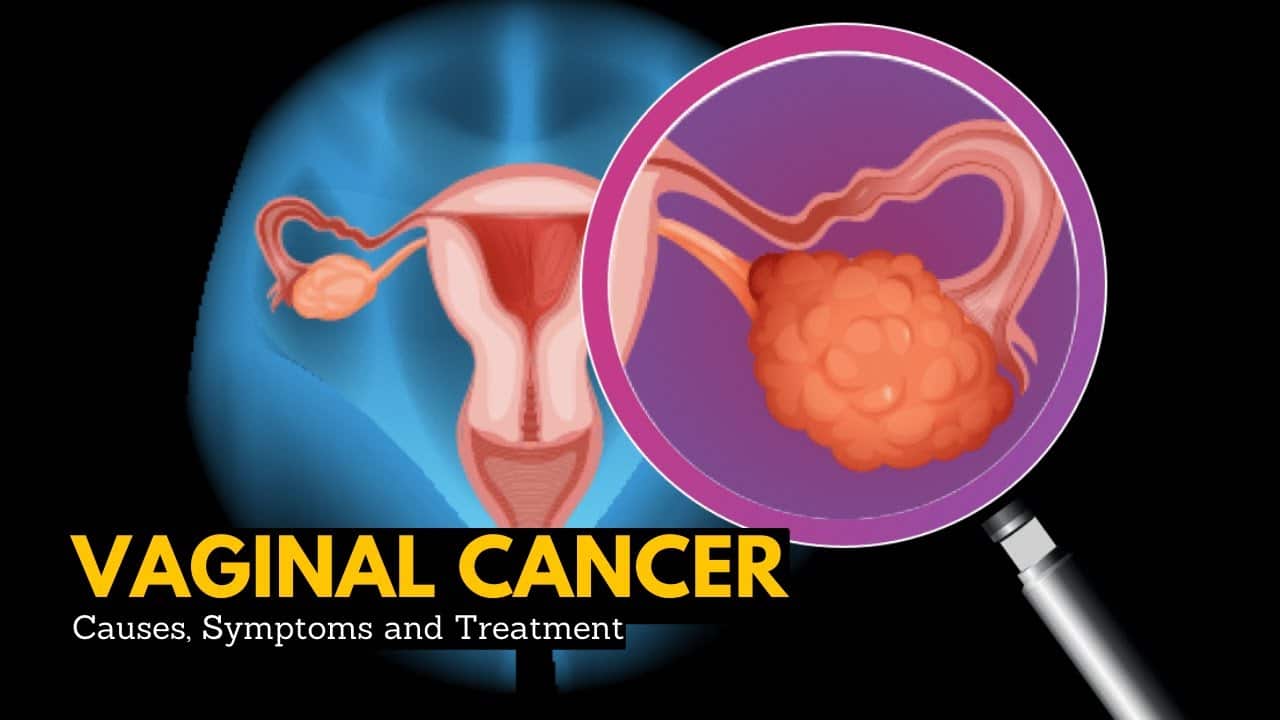
When 28-year-old Jennifer developed abnormal vaginal bleeding and pelvic pain, her gynecologist discovered a vaginal mass during pelvic exam. Biopsy revealed clear cell adenocarcinoma—a rare vaginal cancer typically affecting women in their 60s. “The pathologist asked if my mother took any medications during pregnancy,” Jennifer recalled. “When we checked, Mom found old medical records showing DES prescriptions from 1970. A drug my mother took to prevent miscarriage gave me cancer 28 years later.” Diethylstilbestrol is a synthetic nonsteroidal estrogen, which was widely prescribed to pregnant women between 1938 and 1971 to prevent miscarriages and other pregnancy complications, such as premature delivery. Women whose mothers took DES during pregnancy are at higher risk of developing clear cell adenocarcinoma, a subtype of vaginal cancer. Women exposed to diethylstilbestrol in utero have an increased risk of clear cell adenocarcinoma of the lower genital tract, requiring lifelong cervical and vaginal cancer screening. Studies have demonstrated that women exposed to DES in-utero were 40 times more likely to develop clear cell adenocarcinoma of the cervix or vagina compared with unexposed women, with an incidence of 1 in 1000 in exposed women. Understanding vaginal cancer’s dual epidemiology—elderly women with HPV-related squamous cell cancers and younger “DES daughters” with adenocarcinomas—reveals decades-long consequences of pharmaceutical tragedy alongside contemporary HPV prevention opportunities. JACCUniversity of Rochester Medical Center
A Rare Cancer With Multiple Types
Vaginal cancer represents only 1-2% of gynecologic malignancies—approximately 1,400 U.S. cases annually. According to the CDC, in the U.S., 1,394 women were diagnosed with vaginal cancer in 2022 (the latest year for which data is available) and 438 women died from vaginal cancer in 2022. Tumor must arise primarily in vagina—cancers originating in cervix, vulva, uterus, or other organs metastasizing to vagina don’t qualify. The vagina is the passageway through which fluid passes out of the body during menstrual periods. It is also called the birth canal. The vagina connects the cervix (the opening of the womb, or uterus) and the vulva (the external genitalia). Squamous cell carcinoma (85-90% of cases): arises from vaginal lining squamous epithelium. Strongly linked to HPV infection (particularly HPV-16/18). Squamous cell cancer of the vagina occurs mainly in older women. It can happen at any age, but only a few cases are found in women younger than 40. Almost half of cases occur in women who are 70 years old or older. Appears as ulcerated mass, fungating lesion, or warty growth. Most common location: upper posterior vaginal wall. Adenocarcinoma (5-10%): arises from vaginal glandular cells. Two distinct subtypes: clear cell adenocarcinoma (DES-related, affects younger women 15-40 years); non-clear cell adenocarcinoma (not DES-related, older women 50+ years, worse prognosis than squamous cell). Adenocarcinoma begins in gland cells in your vagina. Clear cell adenocarcinoma is a rare type that’s linked to exposure to a drug called diethylstilbestrol before birth. Melanoma (<3%): arises from pigment-producing melanocytes in vaginal mucosa. Extremely aggressive—worse prognosis than cutaneous melanoma. Appears as darkly pigmented mass or amelanotic (non-pigmented). Sarcoma (<3%): arises from connective tissue/muscle in vaginal wall. Includes leiomyosarcoma (smooth muscle origin), rhabdomyosarcoma (striated muscle—primarily affects children/adolescents). Appears as polypoid mass protruding into vaginal canal. American Cancer Society + 2
The DES Tragedy: A Pharmaceutical Disaster
DES is a hormone drug that was given to pregnant women from the 1940s to early 1970s to prevent miscarriage. The FDA stopped use of this drug in 1971. Diethylstilbestrol (DES): Diethylstilbestrol (DES) is a synthetic form of estrogen prescribed between 1940 and 1971 to prevent pregnancy complications. The promise: synthetic estrogen would prevent miscarriage, premature birth, pregnancy complications by supplementing maternal hormones. Millions of pregnant women prescribed DES 1938-1971 (estimates 5-10 million exposed pregnancies worldwide, 2-3 million in U.S.). The discovery: 1971 study published in New England Journal of Medicine linked DES to clear cell adenocarcinoma in daughters of exposed women. Seven young women (ages 15-22) diagnosed with rare vaginal cancer—all had mothers who took DES during pregnancy. FDA immediately banned DES for pregnancy use (November 1971). The mechanism: DES exposure during first trimester (critical period of reproductive tract development) causes: abnormal development of vaginal/cervical epithelium; vaginal adenosis (glandular tissue where squamous cells should be—found in 30-90% of DES daughters); and structural abnormalities (T-shaped uterus, cervical collars, vaginal ridges). These changes create substrate for malignant transformation years/decades later. There’s about 1 case of this type of cancer in every 1,000 daughters of women who took DES during their pregnancy. This means that about 99.9% of DES daughters do not develop this cancer. DES-related clear cell adenocarcinoma is more common in the vagina than the cervix. The risk appears to be greatest in those whose mothers took the drug during their first 16 weeks of pregnancy. In contrast, in those without DES exposure, clear cell adenocarcinoma of the cervix and vagina is exceedingly rare, with a total of 23 cases identified in the United States from 1931 to 1956. The timing paradox: peak incidence occurred ages 15-25 (median age 19 years at diagnosis). A study at the University of Chicago of clear cell adenocarcinoma registry cases from 1971 to 2014 reported a peak in incidence of DES-related clear cell adenocarcinoma at age 20 years and a second smaller peak at age 42 years. Risk persists lifelong—cases reported into 50s-60s, though declining after age 40. Current status: youngest DES-exposed women now in their 50s (born 1971). Risk continuing but absolute numbers declining as cohort ages. Estimated 1-2 million “DES daughters” in U.S. still requiring lifelong screening. Cleveland Clinic + 4
HPV: The Modern Vaginal Cancer Driver
Human Papillomavirus: HPV infection is a significant risk factor. HPV types 16 and 18 are most strongly linked to vaginal cancer. Risk factors for vaginal cancer include HPV infection, particularly type 16, as well as sexual behavior, chronic vaginitis, and vaginal chemical exposure. The mechanism mirrors cervical cancer: persistent high-risk HPV infection (primarily HPV-16, also HPV-18) integrates into vaginal epithelial cell DNA. Viral oncoproteins E6 and E7 inactivate tumor suppressor genes p53 and Rb. Over 10-30 years, progression occurs: normal epithelium → vaginal intraepithelial neoplasia (VAIN 1, 2, 3) → invasive squamous cell carcinoma. HPV prevalence in vaginal cancers: 60-70% of squamous cell carcinomas HPV-positive (primarily HPV-16). Nearly 100% of high-grade VAIN lesions HPV-positive. Adenocarcinomas (non-DES) occasionally HPV-related. Prevention opportunity: HPV vaccination (Gardasil 9) protects against HPV-16/18 causing majority of vaginal cancers. Ideally administered ages 11-12 before sexual activity; effective through age 26, recommended through age 45 for some. Pap smear screening detects precancerous VAIN lesions allowing treatment before invasive cancer develops—but screening primarily targets cervix, may miss vaginal lesions. Other risk factors: Age: squamous cell carcinoma median age 69 years. Smoking: Smoking doubles the risk of vaginal cancer. Tobacco carcinogens likely contribute to HPV persistence, malignant transformation. Prior cervical cancer/precancer: A history of cervical cancer or precancerous conditions can increase the risk. Shared HPV etiology; field cancerization (entire lower genital tract exposed to carcinogen). Immunosuppression: HIV/AIDS, organ transplant recipients on immunosuppressants—impaired immune surveillance allows HPV persistence, progression. Chronic vaginal irritation: pessary use (for pelvic organ prolapse), untreated vaginal infections, chemical exposures may contribute. Vaginal adenosis: glandular tissue in vagina (present in 30-90% DES daughters but also occurs spontaneously in 5-10% unexposed women)—potential precursor lesion. JACC + 3
Symptoms: Often Silent Until Advanced
Vaginal cancer may not cause symptoms in its early stages. As the cancer grows, symptoms may include: Abnormal Vaginal Bleeding: This can occur after intercourse, between periods, or after menopause. Vaginal Discharge: This may be watery, blood-stained, or have a foul odor. Lump or Mass in the Vagina: A palpable mass may be felt. Pain During Intercourse: The tumor can cause discomfort or pain during sexual activity. You may not have any symptoms of vaginal cancer. If you do, they can include: Other, more common conditions can cause these symptoms. They rarely indicate a condition as serious as vaginal cancer. Abnormal bleeding (most common symptom): postcoital bleeding (after intercourse—tumor friable, bleeds easily with contact), postmenopausal bleeding (any bleeding after menopause warrants investigation), intermenstrual bleeding (between periods), or spotting. Vaginal discharge: watery, blood-tinged, foul-smelling (tumor necrosis, infection). Often dismissed as bacterial vaginosis or yeast infection—persistent discharge despite treatment should prompt examination. Pelvic pain: lower abdominal or pelvic discomfort, may worsen with intercourse, urination, bowel movements. Advanced disease: tumor invading bladder/rectum. Dyspareunia: pain during intercourse—tumor in vaginal canal causes direct trauma, obstruction. Palpable mass: patient or physician may feel firm lump in vagina. Often discovered during routine pelvic exam. Urinary symptoms: frequency, urgency, dysuria, hematuria if tumor invading bladder or compressing urethra. Constipation/rectal bleeding: if tumor invading posterior vaginal wall into rectum. The diagnostic challenge: most symptoms nonspecific, attributed to benign conditions (infections, atrophic vaginitis in postmenopausal women, hemorrhoids). Average delay from symptom onset to diagnosis: 2-6 months. Many cases discovered incidentally during routine pelvic exam or Pap smear showing abnormal cells. JACCnih
Diagnosis: From Pelvic Exam to Biopsy
Pelvic examination: visualization reveals mass, ulceration, discoloration (melanoma). Palpation assesses tumor size, fixation to underlying structures. Speculum exam exposes vaginal walls—important as upper vagina often hidden. Colposcopy: magnified examination of vagina with acetic acid application highlights abnormal areas (acetowhite changes). Guided biopsy of suspicious lesions. Biopsy: definitive diagnosis requires tissue. Punch biopsy under local anesthesia in office for accessible lesions; exam under anesthesia with biopsy for posterior/deep lesions. Cytology: Pap smear may detect abnormal vaginal cells but biopsy required for diagnosis—cytology alone insufficient. Staging workup once diagnosis confirmed: pelvic MRI (best imaging for local tumor extent, parametrial invasion, bladder/rectal involvement); chest CT (lung metastases screening); As with cervical cancer, positron emission tomography/CT has been found to be beneficial in detecting nodal metastasis as well as distant metastasis. One study reported 100% detection of the primary tumor by PET/CT compared with 41% by CT, and 35% detection of enlarged lymph nodes by PET/CT compared with 17% by CT. PET/CT superior to conventional CT for nodal and distant metastasis detection; cystoscopy/proctoscopy if bladder/rectal involvement suspected clinically or on imaging—direct visualization with biopsy confirming invasion. FIGO staging system (clinical staging): Stage I: confined to vaginal wall. Stage II: invades paravaginal tissues but not pelvic sidewall. Stage III: extends to pelvic sidewall. Stage IV: involves bladder mucosa, rectal mucosa, or distant metastases. EBSCO
Treatment: Radiation Dominates, Surgery Limited
Radiation therapy is the treatment of choice in most patients with vaginal cancer, especially in patients with advanced-stage disease. Radiation therapy usually consists of a combination of external beam radiation therapy and brachytherapy. In general, surgery has a limited role in treating vaginal cancer due to the proximity of the cancer to normal tissues such as the bladder, rectum, and urethra. Radiation therapy (primary treatment 70-80% of cases): External beam radiation: treats tumor plus pelvic lymph nodes. Typically 45-50 Gy over 5 weeks. Often combined with concurrent chemotherapy (cisplatin weekly)—radiosensitization improves local control. Brachytherapy (internal radiation): radioactive sources placed directly in/near tumor. High-dose radiation to tumor, sparing surrounding normal tissue. Intracavitary (applicators in vagina) or interstitial (needles through tumor). Critical component—provides boost dose to primary tumor after external beam. Modern intensity-modulated radiation therapy (IMRT) shapes radiation beam conforming to tumor, minimizing bladder/rectum dose—reduces late toxicity (fistulas, strictures, proctitis, cystitis). Surgery (limited role): Stage I small tumors: wide local excision with 1cm margins potentially curative—preserves vaginal function. Stage I upper vagina: radical hysterectomy, upper vaginectomy, pelvic lymphadenectomy if tumor near cervix. Vaginectomy: partial (removing tumor-involved segment) or total (entire vagina). Vaginal reconstruction possible using skin grafts, myocutaneous flaps. Pelvic exenteration: for advanced Stage IV with bladder/rectum involvement or central recurrence after radiation. Removes vagina, uterus, bladder, rectum—creates urinary/fecal diversions. Radical operation, high morbidity—reserved for selected cases. People who had surgery had a 72.1% five-year survival rate compared with 48.9% of those who did not. Similarly, individuals who had radiation therapy had a 59.4% five-year survival rate compared to 52.5% in people who did not. Chemotherapy: limited role. Concurrent chemoradiation (cisplatin) standard for locally advanced disease—radiosensitization only, not systemic treatment. Systemic chemotherapy (cisplatin/5-FU, carboplatin/paclitaxel) for metastatic/recurrent disease—palliative intent, modest benefit. No established effective regimen. EBSCO + 2
Survival: Stage Determines Everything
The relative five-year survival rate is 76% for vaginal cancer found at a localized stage. The relative five-year survival rate is 59% for vaginal cancer with regional spread, through the vaginal wall or nearby lymph nodes. The five-year relative survival rate is 24% for vaginal cancer that has spread to distant organs, such as to the liver, lungs, or bones. In a larger series of patients with vaginal cancer, the 5 year relative survival was 96%, 64–84%, 53–58%, 36%, and 18–36% for stages 0, I, II, III, and IV, respectively. Young women with early-stage DES-related adenocarcinomas treated with either radiation, surgery, or a multimodal approach have good outcomes, with reported 5-year survival in the range of 80%–87%. This is in contrast to non-DES-related adenocarcinomas, which have a worse prognosis. Five-year relative survival by SEER stage (2015-2021 data): Localized (limited to vaginal wall): 76% five-year survival—excellent prognosis if caught early, treated aggressively. Regional (paravaginal tissues, pelvic lymph nodes): 59% five-year survival—requires combined modality treatment, close surveillance. Distant (metastatic to lung, liver, bone): 24% five-year survival—poor prognosis, palliative treatment focus. Additional prognostic factors: Tumor size: Tumor size >4 cm is associated with decreased local control of disease and worsened overall survival. Other factors that negatively affect prognosis include tumor size >4 cm, older age, and tumor location outside of the upper third of the vagina. Tumors >4cm significantly worse outcomes than smaller lesions. Age: older patients (>70 years) worse survival—comorbidities, treatment tolerance, advanced stage at diagnosis. Location: lower third vagina worse prognosis than upper third—proximity to vulva, more difficult radiation targeting. Histology: In general, adenocarcinomas have a worse prognosis than squamous cell carcinoma. Histological subtype also affects prognosis as adenocarcinomas have a worse prognosis than squamous cell carcinoma. Melanoma worst prognosis (median survival 12-24 months even with treatment). HPV status: high-risk HPV-positive tumors paradoxically better prognosis—more radiosensitive, similar to cervical cancer. The DES daughter exception: young women with DES-related clear cell adenocarcinoma have relatively favorable prognosis (80-87% five-year survival) when diagnosed early, treated appropriately—better than non-DES adenocarcinomas or squamous cell carcinomas. Risk Factors for Vaginal Cancer | American Cancer Society +2 + 2
Screening and Prevention: The DES Daughter Protocol
Standard screening (all women): annual pelvic exam with careful vaginal inspection; Pap smear includes vaginal walls, not just cervix (many practitioners sample cervix only); and HPV vaccination recommended through age 45 for those not previously vaccinated. DES daughter screening (enhanced protocol): Women exposed to diethylstilbestrol in utero have an increased risk of clear cell adenocarcinoma of the lower genital tract, requiring lifelong cervical and vaginal cancer screening. Women who are exposed should undergo more frequent screenings at the physician’s and patient’s discretion. Annual pelvic exam starting age 14-15 or onset of menstruation; Pap smear of cervix AND four-quadrant vaginal sampling (clear cell adenocarcinoma can arise anywhere in vagina); palpation of entire vaginal length (tumors may be submucosal, not visible); colposcopy with iodine staining (Lugol’s iodine highlights abnormal areas)—some experts recommend every 1-2 years; and continued screening throughout life (risk persists—no age to discontinue). The challenge: most DES daughters now 50s-60s; many providers unfamiliar with DES history or specialized screening needs. DES daughters should see gynecologists knowledgeable about DES-related abnormalities. Prevention strategies: HPV vaccination (prevents majority of squamous cell carcinomas); smoking cessation (doubles risk); prompt treatment of persistent vaginal infections, discharge; and regular pelvic exams allowing early detection before symptoms develop. JACCJohns Hopkins Medicine
Frequently Asked Questions
Q1: My mother took DES during pregnancy in 1968. What’s my actual cancer risk and what screening do I need?
Your lifetime risk of developing vaginal/cervical clear cell adenocarcinoma is approximately 1 in 1,000 (0.1%)—40 times higher than unexposed women but still meaning 99.9% of DES daughters never develop this cancer. The cumulative risk of cancer as 1 in 750 up to the age of 50. Peak risk occurred ages 15-25 (median 19 years); at age 56-57 now, your risk substantially lower than during young adulthood but not zero—cases reported into 60s. Recommended screening: annual gynecologic exam with four-quadrant vaginal Pap smear (sampling cervix plus all vaginal walls—not just cervix); careful palpation entire vaginal length checking for submucosal masses; colposcopy every 1-2 years with iodine staining optional but helpful; and breast exams (DES exposure also linked to slightly increased breast cancer risk). Find gynecologist familiar with DES screening protocols—many younger providers never encountered DES patients. Additional DES-related issues beyond cancer: structural abnormalities (T-shaped uterus, cervical abnormalities) may have caused fertility problems, pregnancy complications; vaginal adenosis (glandular tissue in vagina) benign but requires monitoring; increased risk cervical dysplasia/cancer (not just vaginal). Don’t panic—absolute risk remains low, but lifelong vigilance appropriate. University of Rochester Medical Center
Q2: I’m 72 with postmenopausal bleeding. My doctor says it’s probably atrophic vaginitis but wants biopsy. How worried should I be about vaginal cancer?
Postmenopausal bleeding always requires investigation—but vaginal cancer remains uncommon even with this symptom. Likely causes (in descending order): endometrial pathology (polyps, hyperplasia, endometrial cancer—most common), atrophic vaginitis (thinning vaginal tissues bleed easily), cervical polyps/lesions, vulvar lesions, and vaginal cancer (rare but must be excluded). Your doctor’s approach is correct: pelvic exam visualizing bleeding source; endometrial biopsy (if uterus intact—excludes endometrial cancer); and vaginal biopsy if suspicious lesions visible. Risk factors increasing vaginal cancer likelihood in your case: age 72 (squamous cell carcinoma median age 69); history of abnormal Pap smears, cervical dysplasia/cancer; smoking history; HPV infection; and immunosuppression. Even with risk factors, atrophic vaginitis remains more likely—but biopsy provides definitive answer. If vaginal cancer diagnosed: likely squamous cell carcinoma (85-90%); staging determines treatment—localized disease very treatable with radiation; and prognosis depends on stage—76% five-year survival for localized disease. Don’t delay biopsy due to fear—early detection dramatically improves outcomes. Postmenopausal bleeding investigated promptly has better prognosis than symptoms ignored until advanced.
Q3: I was just diagnosed with Stage II vaginal squamous cell carcinoma. What does treatment involve and what’s my prognosis?
Stage II means tumor has invaded paravaginal tissues but hasn’t reached pelvic sidewall—locally advanced but potentially curable. Standard treatment: concurrent chemoradiation (external beam radiation to pelvis plus weekly cisplatin chemotherapy for radiosensitization) 5-6 weeks; brachytherapy boost (internal radiation to tumor) 1-2 weeks after external beam completes; and total treatment duration approximately 7-8 weeks. Side effects during treatment: fatigue (cumulative, worsens over weeks); vaginal discharge (tumor breakdown, inflammation); diarrhea, bladder irritation (radiation affects nearby rectum, bladder); skin changes (groin, perineum—like sunburn); and temporary reduction in white blood cells, platelets (chemotherapy effect). Long-term effects: vaginal stenosis (narrowing, shortening from fibrosis)—vaginal dilator use essential preventing complete closure; sexual dysfunction (decreased lubrication, dyspareunia)—manageable with lubricants, counseling; chronic proctitis, cystitis (5-15%)—occasional rectal bleeding, urinary urgency; and premature menopause if ovaries in radiation field (depending on age). Prognosis: The relative five-year survival rate is 59% for vaginal cancer with regional spread. Stage II falls into this category—approximately 53-58% five-year survival depending on tumor size, exact extent. Factors improving your prognosis: tumor <4cm; HPV-positive (if squamous cell—better radiosensitivity); completion of full radiation course (don’t skip treatments—critical for cure); and younger age, good performance status. Follow-up: pelvic exams every 3 months years 1-2, every 6 months years 3-5, annually thereafter; imaging (CT/MRI) as needed checking for recurrence; and vaginal dilator use 2-3 times weekly lifelong preventing stenosis. While 59% five-year survival isn’t as favorable as early-stage, majority of women with Stage II disease achieve long-term survival with aggressive treatment. American Cancer Society
Q4: Can vaginal cancer be prevented?
Partially, yes—particularly for HPV-related squamous cell carcinomas. Prevention strategies: HPV vaccination: Gardasil 9 protects against HPV-16/18 causing 70%+ of vaginal squamous cell cancers. Ideally administered ages 11-12 before sexual activity; effective through age 26; recommended for catch-up vaccination through age 45 in some. Vaccination prevents most vaginal cancers (along with cervical, vulvar, oropharyngeal, anal cancers). Smoking cessation: smoking doubles vaginal cancer risk. Quitting reduces risk over time. Regular screening: annual pelvic exams allow detection of precancerous VAIN lesions treated before invasive cancer develops. Pap smears (including vaginal walls) detect abnormal cells. Prompt treatment of infections: chronic vaginal inflammation (untreated bacterial vaginosis, yeast infections) may contribute to cancer risk—though evidence weaker than other factors. Safe sexual practices: limiting partners, condoms (reduce but don’t eliminate HPV transmission). DES exposure: can’t be prevented now (drug banned 1971) but DES daughters should follow enhanced screening detecting cancers early when most curable. What can’t be prevented: age (squamous cell carcinoma risk increases with age); prior cervical cancer/precancer (shared HPV etiology); and immunosuppression (HIV, transplant medications impair immune surveillance). Bottom line: HPV vaccination prevents majority of future vaginal cancers. For current adults beyond vaccination window, regular screening best prevention—detecting precancerous lesions or early cancers when highly curable.
Q5: I had radiation for cervical cancer 15 years ago. Does this increase my vaginal cancer risk?
Yes, modestly. Prior pelvic radiation increases secondary vaginal cancer risk through two mechanisms: Radiation-induced malignancy: ionizing radiation damages DNA in normal tissues. Latency period typically 10-30 years. Risk proportional to radiation dose received. Secondary cancers in radiation field include vaginal, vulvar, bladder, rectal cancers. Absolute risk remains low—estimated 1-2% developing secondary malignancy over 20-30 years. HPV field effect: if cervical cancer was HPV-related (most are), entire lower genital tract exposed to same HPV strain. Risk of developing second primary in vagina, vulva even without radiation. Radiation may impair local immune surveillance facilitating HPV persistence. Your surveillance protocol: annual pelvic exam with careful vaginal inspection; Pap smear including vaginal walls (not just cervix, since you had cervical cancer); colposcopy if abnormalities detected; low threshold for biopsy of suspicious areas; and report any symptoms (bleeding, discharge, pain) promptly—don’t dismiss as radiation effects. The cervical cancer treatment paradox: radiation cured your cervical cancer but created small long-term secondary cancer risk. Overall benefit massively outweighs risk—without treatment, cervical cancer would have progressed. The key: lifelong vigilance with regular screening allowing early detection if second primary develops. Most radiation patients never develop secondary cancers; those who do typically catch them early due to close surveillance.
Disclaimer
This article adapts publicly available information from reputable cancer research organizations and medical databases. This content is for informational and educational purposes only and does not constitute medical advice. ObserverVoice.com is a news and information platform — not a healthcare provider. Decisions about vaginal cancer screening, diagnosis, and treatment should be made in consultation with qualified physicians, gynecologists, and oncologists who can evaluate your individual symptoms, risk factors, and health status. If you have symptoms concerning for vaginal cancer or are a DES daughter requiring specialized screening, please consult with your healthcare team promptly.
References
- OncoDaily. Vaginal Cancer: New insights in 2024. https://oncodaily.com/oncolibrary/cancer-types/vaginal-cancer-74679
- Cleveland Clinic. Vaginal Cancer: Causes, Symptoms, Types & Treatment. https://my.clevelandclinic.org/health/diseases/15579-vaginal-cancer
- American Cancer Society. Risk Factors for Vaginal Cancer. https://www.cancer.org/cancer/types/vaginal-cancer/causes-risks-prevention/risk-factors.html
- PMC. Post-menopausal vaginal and cervical cancer risk related to in utero diethylstilbestrol exposure. https://pmc.ncbi.nlm.nih.gov/articles/PMC10311286/
- PMC. Updates in the treatment of vaginal cancer. https://pmc.ncbi.nlm.nih.gov/articles/PMC8921584/
Observer Voice is the one stop site for National, International news, Sports, Editor’s Choice, Art/culture contents, Quotes and much more. We also cover historical contents. Historical contents includes World History, Indian History, and what happened today. The website also covers Entertainment across the India and World.

