

Normal Pressure Hydrocephalus: The Treatable Dementia Mimic

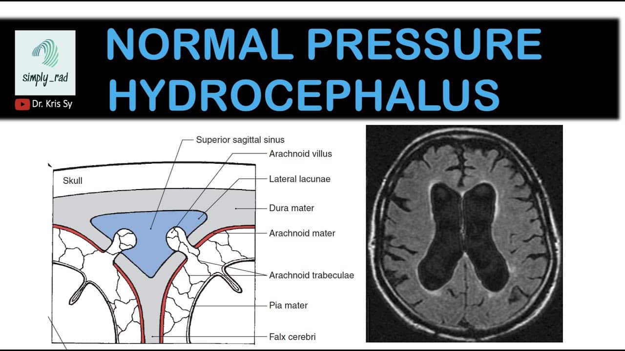
Imagine a person developing cognitive decline, memory loss, and slowed thinking—seemingly classic dementia. Simultaneously, their gait becomes slow and unsteady. They shuffle when walking. They fall frequently. Additionally, they develop urinary incontinence. Brain imaging shows enlarged ventricles. However, unlike typical dementia, this condition can be partially or even completely reversed through a simple surgical procedure—ventriculoperitoneal shunting. This is normal pressure hydrocephalus—a rare but treatable condition where cerebrospinal fluid accumulation in the brain’s ventricles causes a distinctive triad of symptoms that mimics dementia but can improve dramatically with appropriate treatment. Normal pressure hydrocephalus, commonly abbreviated as NPH, is a neurological disorder characterized by enlargement of the brain’s ventricles despite normal cerebrospinal fluid pressure. The ventricles are fluid-filled cavities within the brain. Normally, cerebrospinal fluid (CSF) is produced in the ventricles by the choroid plexus. The fluid flows through the ventricular system and circulates around the brain and spinal cord. The fluid is reabsorbed into the bloodstream through arachnoid granulations. In normal pressure hydrocephalus, the balance between CSF production and absorption is disrupted. CSF accumulates in the ventricles. The ventricles enlarge. Despite the ventricular enlargement, the intracranial pressure remains normal or only mildly elevated—hence the name “normal pressure.” The ventricular enlargement directly damages neurons. The stretching distorts neural pathways. The pressure on white matter tracts disrupts neuronal communication. The result is the distinctive triad of symptoms: cognitive decline, gait disturbance, and urinary incontinence. Normal pressure hydrocephalus accounts for approximately 5 to 10 percent of dementia cases. However, many cases go undiagnosed. Patients are mistakenly diagnosed with Alzheimer’s disease or Parkinson’s disease. The misdiagnosis is unfortunate because, unlike Alzheimer’s disease, normal pressure hydrocephalus can improve or even partially reverse with appropriate treatment. What makes normal pressure hydrocephalus particularly important is its treatability. Ventriculoperitoneal shunting diverts CSF away from the brain. The shunt allows CSF to drain into the peritoneal cavity where it is absorbed. The shunting relieves ventricular pressure. The ventricular enlargement decreases. Neural compression is relieved. Cognitive function can improve. Gait can normalize. Continence can be restored. In this comprehensive article, we will explore what normal pressure hydrocephalus is, understand how CSF accumulation damages cognition and gait, recognize the distinctive symptom triad, learn about disease mechanisms, understand diagnostic methods, explore treatment options, and discover why this dementia mimic is so important to identify.
Understanding Cerebrospinal Fluid and Ventricular System
Before we explore normal pressure hydrocephalus, we need to understand cerebrospinal fluid production, circulation, and absorption. The brain floats in cerebrospinal fluid. The fluid provides buoyancy reducing brain weight. The brain weighs approximately 1400 grams. In air, the brain’s effective weight is approximately 50 grams—supported by CSF buoyancy. The CSF provides mechanical protection. The fluid cushions the brain against trauma. The CSF provides immune function. White blood cells in CSF patrol for infection. The CSF removes waste products from the brain. Metabolic waste from neurons is cleared by the glymphatic system. The glymphatic system relies on CSF circulation. CSF is produced in the ventricles. The choroid plexus lines the ventricles. The choroid plexus is specialized tissue producing CSF. CSF production is approximately 400 to 500 milliliters per day. This enormous production rate—the entire ventricular system is completely refreshed multiple times daily. The ventricles are fluid-filled cavities within the brain. The lateral ventricles are paired cavities in the cerebral hemispheres. The third ventricle is a midline cavity below the lateral ventricles. The fourth ventricle is a cavity in the brainstem. The ventricles communicate through narrow passages called foramina. CSF flows from the lateral ventricles through the foramen of Monro into the third ventricle. CSF flows from the third ventricle through the cerebral aqueduct into the fourth ventricle. CSF exits the fourth ventricle through the foramen of Luschka and foramen of Magendie into the subarachnoid space. The subarachnoid space surrounds the brain and spinal cord. CSF circulates through the subarachnoid space. The CSF bathes the brain and spinal cord. CSF is reabsorbed into the bloodstream. Arachnoid granulations are specialized structures in the dura mater. The granulations project into dural venous sinuses. CSF passes through granulations into venous blood. The reabsorption is proportional to intracranial pressure. Normal intracranial pressure is approximately 10 to 15 mmHg. Higher pressure increases reabsorption. CSF pressure and volume are tightly regulated. The balance between production (400-500 mL/day) and reabsorption maintains normal pressure and volume. In normal pressure hydrocephalus, this balance is disrupted. CSF production continues normally. CSF reabsorption is impaired. CSF accumulates. Ventricular pressure rises acutely causing distension. Eventually, pressure normalizes despite ventricular enlargement. The mechanism for pressure normalization is incompletely understood but might involve increased reabsorption as ventricles enlarge. Understanding CSF physiology helps explain why normal pressure hydrocephalus develops and why shunting helps.
What is Normal Pressure Hydrocephalus?
Normal pressure hydrocephalus is a neurological disorder where impaired CSF absorption leads to ventricular enlargement and progressive neurological dysfunction despite normal intracranial pressure. The disease is characterized by the clinical triad: cognitive decline, gait disturbance, and urinary incontinence. However, not all patients have all three features. The pattern varies. What causes impaired CSF absorption in normal pressure hydrocephalus is incompletely understood. Idiopathic normal pressure hydrocephalus has no identified cause. The cause is unknown. Arachnoid granule dysfunction might reduce CSF reabsorption. The granules might be blocked by debris. The granules might have reduced absorptive capacity. The mechanism is speculative. Secondary normal pressure hydrocephalus results from identifiable causes. Prior subarachnoid hemorrhage—bleeding in the subarachnoid space—damages arachnoid granules. Post-traumatic hydrocephalus develops after head injury. Head trauma damages CSF pathways. Arachnoid granule scar tissue blocks CSF reabsorption. Meningitis—infection of brain membranes—causes inflammation. The inflammation damages arachnoid granules. Post-infectious hydrocephalus results from granule damage. Tumor or mass blocking CSF flow. Obstruction prevents normal circulation. Pressure builds. Ventricular dilation develops. This is obstructive hydrocephalus, which is technically different from normal pressure hydrocephalus but can present similarly. Normal pressure hydrocephalus affects older adults predominantly. The disease is rare before age 60. The disease is slightly more common in men. The disease is rare in women. The onset is insidious. Symptoms develop gradually over months to years. The gradual onset makes diagnosis difficult. The insidious presentation allows significant brain damage before diagnosis. Early symptoms are subtle. Cognitive changes are minimal initially. Gait changes are subtle. Incontinence might be mild. Over time, symptoms progress. Cognitive decline becomes more obvious. Gait becomes more impaired. Incontinence worsens. The progressive nature parallels dementia. The similarity to dementia causes misdiagnosis. Many patients are labeled with Alzheimer’s disease. The misdiagnosis delays appropriate treatment. Shunting might be considered late—after significant irreversible damage has occurred. The timing of shunting influences outcomes. Early shunting before irreversible damage produces better results. Late shunting after years of ventricular enlargement produces less improvement. Some damage becomes irreversible. The importance of early diagnosis cannot be overstated.
Recognizing the Classic Triad: Cognitive, Gait, and Continence Changes
Normal pressure hydrocephalus causes a distinctive triad of symptoms. Recognizing the triad helps diagnosis. Cognitive decline is the first symptom often. Memory becomes impaired. Recent memory is affected—difficulty remembering recent events. Long-term memory might be relatively preserved. Concentration becomes difficult. Attention becomes impaired. Processing becomes slower. Executive function becomes impaired. Planning and organization become difficult. Decision-making becomes difficult. The cognitive decline resembles dementia. Patients are often misdiagnosed with Alzheimer’s disease. The cognitive decline is prominent. Personality might change. Apathy develops. Motivation decreases. The person becomes passive. Mood changes develop. Depression is common. Mood lability—rapid mood changes—occurs. Emotional blunting might occur. The person appears emotionally flat. These mood and personality changes resemble dementia or depression. The cognitive decline progresses. Without treatment, cognitive decline worsens progressively. Dementia becomes advanced. The cognitive decline can become severe and irreversible. Gait disturbance is often the most distinctive symptom. The gait is classically described as “magnetic” or “marching.” The feet appear stuck to the floor. Walking requires great effort. The person must consciously think about walking. The gait becomes slow. The shuffling gait resembles Parkinson’s disease. However, unlike Parkinson’s disease, tremor is absent. Unlike Parkinson’s disease, rigidity is not prominent. Unlike Parkinson’s disease, the patient does not have difficulty initiating movement (bradykinesia) in a parkinsonian sense. The gait disturbance is distinctive. The feet move short distances. The person shuffles. The feet feel heavy. The stride length is reduced. Walking becomes laborious. Falls become frequent. The person becomes increasingly unsteady. Balance becomes impaired. The gait disturbance is often the symptom that brings patients to medical attention. The gait becomes so abnormal that disability is obvious. Urinary incontinence is the third classic symptom. The person loses bladder control. Urgency develops—the urge to urinate becomes frequent and urgent. Urge incontinence develops—the person cannot hold urine and leaks. The incontinence develops gradually. Initially mild—occasionally leaking. Progressively worsens. Severe incontinence requiring diapers develops. The incontinence resembles overactive bladder. However, urodynamic testing might reveal normal bladder function. The incontinence results from impaired central control of bladder function. The dilated ventricles affect pathways controlling micturition. The person loses normal inhibition of bladder contraction. The incontinence can develop after cognitive decline and gait disturbance. In some patients, incontinence is the predominant symptom. The classic triad—cognitive decline, gait disturbance, and urinary incontinence—is distinctive. However, not all patients have all three features. Some patients have cognitive decline and gait disturbance without incontinence. Some have prominent gait disturbance with mild cognitive changes. The incomplete presentation causes diagnostic confusion. Recognizing any component of the triad should prompt brain imaging. The combination strongly suggests normal pressure hydrocephalus. The imaging can confirm or exclude diagnosis.
Understanding Ventricular Enlargement and Neurological Dysfunction
Understanding how enlarged ventricles cause neurological dysfunction helps explain the symptom pattern. Ventricular enlargement causes direct mechanical effects. The enlarged ventricles take up space. Adjacent brain tissue is compressed. The compression is gentle—not acute like a tumor. However, chronic compression gradually damages tissue. White matter tracts are stretched. The stretching impairs axonal function. Myelin—the insulation on axons—is damaged. The damaged white matter tracts disrupt communication between brain regions. The disrupted communication causes cognitive and motor dysfunction. Memory-related pathways are stretched. Memory function is impaired. Motor pathways are stretched. Gait control is impaired. Micturition pathways are disrupted. Continence is lost. The specific symptoms reflect which pathways are stretched. Periventricular white matter is affected most. The white matter surrounding the ventricles is stretched maximally. The corpus callosum—the structure connecting the two hemispheres—can be disrupted. Interhemispheric communication is impaired. Cognitive and motor coordination suffer. The anterior and posterior horns of the lateral ventricles enlarge. The frontal horn enlargement compresses frontal white matter. Executive function and motor control suffer. The gait center is located in medial frontal lobe. Stretching of this region causes gait dysfunction. The temporal horn enlargement compresses temporal structures. Memory systems are affected. Memory impairment results. The posterior horn enlargement compresses parietal and occipital white matter. Visuospatial and cognitive functions suffer. The differential stretching of different ventricular regions explains the variable symptom presentation. The degree of ventricular enlargement varies. Mild enlargement causes subtle symptoms. Severe enlargement causes profound symptoms. The relationship between ventricular size and symptom severity is not linear. Some patients have minimal symptoms despite large ventricles. Others have severe symptoms with modest enlargement. The variability in symptom severity reflects individual differences in tolerance or compensation. Intracranial compliance—how the skull accommodates changes—influences symptoms. Patients with poor compliance develop symptoms earlier. The timing of symptom development influences severity. Acute ventricular enlargement causes severe symptoms. Chronic gradual enlargement allows some neuronal adaptation. The chronic process allows some compensation. However, compensation is incomplete. Progressive dysfunction develops. The ventricular enlargement and white matter stretching are potentially reversible through shunting. If shunting relieves the stretch, white matter function can recover. Earlier shunting before irreversible damage increases recovery potential. This reversibility makes early diagnosis and treatment crucial.
Diagnosis: Recognizing the Dementia Mimic
Diagnosing normal pressure hydrocephalus requires clinical suspicion and appropriate imaging. Clinical history is crucial. Doctors should ask about the symptom triad. When did symptoms start? What changed—memory, gait, or continence? Have all three symptoms developed or only some? Family history might help—normal pressure hydrocephalus is occasionally familial. Prior head trauma, meningitis, or subarachnoid hemorrhage increases risk. Prior medical events might trigger investigation. Physical examination documents findings. Neurologic examination. Cognitive testing—Mini-Cog or Montreal Cognitive Assessment. Gait assessment is crucial. The distinctive “magnetic” gait is characteristic. The person walks slowly with shuffling gait. The feet drag. The feet feel stuck. The gait appears stuck in wet concrete. The gait is distinctive. Movement assessment. Rigidity assessment—usually absent or minimal. Tremor assessment—should be absent. Bradykinesia—usually absent. Postural stability assessment. Cranial nerve assessment. Reflexes—usually normal. Balance assessment. Incontinence history. The combination of cognitive impairment, distinctive gait, and incontinence raises suspicion. Brain imaging is essential. MRI brain shows enlarged ventricles. Ventricular enlargement is the key finding. The lateral ventricles are enlarged. The third ventricle is enlarged. The fourth ventricle is usually normal. Periventricular hyperintensities appear on T2 and FLAIR sequences. White matter hyperintensities in the periventricular region. These represent transependymal CSF flow—CSF leaking through ventricular walls. The hyperintensities are characteristic of hydrocephalus. Cortical sulci might be normal or only mildly enlarged. This is important. In Alzheimer’s disease, cortical sulci are prominently enlarged from brain atrophy. In normal pressure hydrocephalus, sulci are normal or only mildly enlarged. The preserved sulci with enlarged ventricles is distinctive. This pattern—enlarged ventricles with normal sulci—is characteristic of normal pressure hydrocephalus. CT brain shows ventricular enlargement. CT is less sensitive for white matter changes than MRI. However, CT clearly shows enlarged ventricles. Lumbar puncture obtains CSF. Opening pressure is measured. Normal pressure hydrocephalus typically has normal or only mildly elevated pressure (less than 25 cmH2O). This normal pressure despite ventricular enlargement is pathognomonic. Elevated protein and elevated cell count suggest secondary hydrocephalus from infection or bleeding. Normal chemistry supports idiopathic normal pressure hydrocephalus. CSF tap test—removing 40-50 mL of CSF—sometimes causes acute symptom improvement. Symptom improvement after CSF removal predicts shunt responsiveness. However, the tap test is imperfect. Absence of improvement does not exclude normal pressure hydrocephalus. Gait improvement after CSF removal is more predictive than cognitive improvement. Infusion test—slowly infusing CSF while monitoring pressure—assesses CSF absorption. Reduced CSF absorption indicates normal pressure hydrocephalus. However, the test is complex and not universally performed. Dynamic MRI—imaging while CSF flow is assessed—might help predict shunt response. However, it is not routinely performed. The diagnosis of normal pressure hydrocephalus is based on: clinical triad plus ventricular enlargement on imaging plus normal or mildly elevated CSF pressure. Some diagnostic uncertainty is expected. The condition mimics dementia. Imaging might not be perfectly distinctive. CSF pressure might be borderline. The diagnosis is sometimes “probable normal pressure hydrocephalus.” The certainty increases when all three components are clearly present.
Treatment: Ventriculoperitoneal Shunting and Recovery
Treatment for normal pressure hydrocephalus is ventriculoperitoneal (VP) shunting. The shunt diverts CSF away from the brain. A small tube is placed in the lateral ventricle. The tube is threaded under the skin. The tube drains into the peritoneal cavity (abdominal cavity). A valve regulates flow. The valve allows CSF to drain when pressure exceeds the valve setting. The valve prevents reverse flow. CSF is continuously drained. The ventricles decrease in size. Periventricular white matter is decompressed. Neural function improves. The shunt relieves ventricular pressure. The ventricular enlargement decreases. The periventricular hyperintensities might improve. The white matter recovers. Neurological function improves. Symptom improvement follows shunting. Cognitive function improves. Memory improves. Processing speed increases. Gait normalizes. The shuffling gait becomes more normal. Walking becomes easier. Balance improves. Falls decrease. Continence improves. Incontinence resolves or dramatically improves. The symptom improvement is often dramatic. Some patients describe cognitive and functional normalization. The improvement reflects white matter decompression and recovery. Timing of shunting is crucial. Early shunting before irreversible white matter damage produces better results. Late shunting after years of ventricular enlargement produces less improvement. Some white matter damage becomes irreversible. Axons severely damaged might not recover. Demyelination from prolonged stretching might be permanent. Early diagnosis and early treatment optimize outcomes. The delay in diagnosis is problematic. Many patients suffer for years with unrecognized normal pressure hydrocephalus. They are treated for Alzheimer’s disease. They are treated for Parkinson’s disease. They are treated for depression. Meanwhile, their ventricular enlargement continues. Their white matter gradually degenerates. Years pass before correct diagnosis. By then, some damage is permanent. Outcome is suboptimal. Shunt complications occur in approximately 30 to 40 percent of patients. Shunt malfunction—the shunt stops working. Ventricular re-enlargement occurs. Symptoms recur. Shunt revision is necessary. Shunt infection—bacteria colonize the shunt. Meningitis develops. The infected shunt must be removed. Antibiotics treat infection. A new shunt is placed after sterilization. Over-drainage—too much CSF is drained. Subdural hematomas develop—bleeding between membranes. Headaches develop. The valve setting must be adjusted. Under-drainage—too little CSF is drained. Symptoms persist. The valve pressure must be decreased. Shunt failure is the most common complication. Shunts fail at a rate of approximately 10 to 15 percent per year. Multiple shunt revisions are often necessary over lifetime. Multiple surgeries carry cumulative risks. However, despite complications, shunting dramatically improves quality of life for many patients. The improvement justifies the risks.
Living with Normal Pressure Hydrocephalus: Pre and Post-Treatment
Living with normal pressure hydrocephalus before treatment diagnosis is difficult. Cognitive decline causes disability. Memory loss causes functional impairment. Gait disturbance causes falls and injuries. Incontinence causes social embarrassment and burden. The person becomes increasingly dependent. The misdiagnosis as Alzheimer’s disease or Parkinson’s disease is common. Treatment is directed at the wrong condition. Levodopa is given for presumed Parkinson’s—ineffective. Cognitive enhancers are given for Alzheimer’s—ineffective or harmful. The person deteriorates despite treatment. The misdiagnosis causes suffering. The delay in correct diagnosis is harmful. Years might pass before brain imaging clearly shows the problem. The imaging must be properly interpreted. The enlarged ventricles must be recognized. The normal sulci must be appreciated. The periventricular hyperintensities must be noted. Some radiologists miss the findings. The diagnosis can be delayed despite imaging. Once diagnosed, evaluation continues. Lumbar puncture is performed. CSF pressure is measured. CSF analysis is done. CSF tap test might be performed. The diagnostic confirmation takes time. The patient waits. The ventricular enlargement continues. The white matter damage accumulates. Finally, shunt surgery is scheduled. The surgical intervention. General anesthesia is used. A small burr hole is made in the skull. A catheter is placed in the lateral ventricle. The catheter is tunneled under skin. A valve is placed under the clavicle. The catheter is tunneled to the abdomen. A large hole is made in the peritoneum. The catheter is placed in the peritoneal cavity. Sutures close the incisions. The shunt system is in place. CSF now drains from ventricles into abdomen. Recovery from surgery. The surgical wounds heal. The shunt begins functioning. CSF drains continuously. Ventricular enlargement decreases. Neural decompression occurs. Symptom improvement begins. The improvement might occur immediately. The improvement might occur gradually over weeks. The improvement might reach maximum within months. Some patients recover dramatically. Cognitive function normalizes. Gait normalizes. Continence is restored. Life normalizes. Other patients improve partially. Some cognitive dysfunction persists. Some gait abnormality remains. Some incontinence persists. But improvement is significant. Quality of life substantially improves. Post-treatment management. Shunt surveillance. Regular imaging monitors shunt function. Ventricular size is tracked. Periventricular changes are monitored. Clinical assessment monitors symptom status. Cognitive reassessment. Gait reassessment. Continence status. Shunt dysfunction signs prompt evaluation. Symptom recurrence—cognitive decline, gait worsening. Headache, nausea, vomiting suggest shunt malfunction. Immediate evaluation is necessary. Shunt revision might be necessary. Imaging shows ventricular re-enlargement. The shunt is revised. Long-term follow-up continues. Multiple shunt revisions over lifetime are common. Each revision carries surgical risk. But the benefit of shunt function justifies the risks. Quality of life with functional shunt is dramatically better than without. Living with normal pressure hydrocephalus post-treatment is dramatically improved. The cognitive decline is halted or reversed. The gait normalizes. Incontinence improves. The person regains independence. Life resumes normal patterns. Employment might resume. Social engagement normalizes. Family relationships improve. The transformation from disabled to functional is remarkable for many patients.
Frequently Asked Questions (FAQs)
Q1: Can normal pressure hydrocephalus be confused with Alzheimer’s disease?
Yes, normal pressure hydrocephalus is frequently misdiagnosed as Alzheimer’s disease. Both cause cognitive decline and dementia. However, normal pressure hydrocephalus also causes the distinctive gait disturbance and incontinence. Alzheimer’s disease does not typically cause these features. The gait and incontinence help differentiate. However, without recognizing these features or obtaining brain imaging, the conditions are easily confused. Many patients are misdiagnosed.
Q2: Is normal pressure hydrocephalus surgical emergency?
Normal pressure hydrocephalus is not an acute emergency like acute hydrocephalus from tumor or hemorrhage. However, it is a chronic progressive condition. The earlier shunting is performed, the better the outcome. The longer the delay, the more white matter damage accumulates. Some damage becomes irreversible. The condition should be treated urgently when diagnosed—within weeks rather than months. Delays allow preventable deterioration.
Q3: What is the success rate of shunt surgery?
Approximately 50 to 70 percent of appropriately selected normal pressure hydrocephalus patients show significant improvement after shunting. Some patients have dramatic cognitive and functional recovery. Others improve partially. Approximately 20 to 30 percent have minimal improvement. The reasons for variable response are incompletely understood. Presence of other dementia pathology (Alzheimer’s disease) reduces response. Duration of symptoms before shunting affects response. Earlier shunting produces better results.
Q4: Can normal pressure hydrocephalus return after successful shunt placement?
Yes, normal pressure hydrocephalus can recur. Shunt malfunction is common. Approximately 10 to 15 percent of shunts fail per year. Shunt failure allows ventricular re-enlargement. Symptoms recur. Shunt revision is necessary. Over a lifetime, multiple shunt revisions are often needed. Each revision carries surgical risk but maintains function.
Q5: How can normal pressure hydrocephalus be distinguished from Parkinson’s disease?
Normal pressure hydrocephalus gait resembles Parkinson’s disease gait. However, important differences exist. Parkinson’s gait has reduced arm swinging and stooped posture. Parkinson’s tremor is characteristic. Parkinson’s has rigidity and bradykinesia. Normal pressure hydrocephalus lacks these features. Normal pressure hydrocephalus has cognitive decline and incontinence—not typical of Parkinson’s. Brain imaging showing enlarged ventricles with normal sulci supports NPH diagnosis.
Key Takeaways
Normal pressure hydrocephalus is a treatable neurological disorder caused by impaired CSF absorption leading to ventricular enlargement. NPH accounts for 5 to 10 percent of dementia cases but is frequently undiagnosed. The classic triad includes cognitive decline, gait disturbance, and urinary incontinence. Cognitive decline resembles dementia—memory loss, slowed thinking, executive dysfunction. Gait disturbance is distinctive—slow, shuffling “magnetic” gait. Urinary incontinence develops from impaired micturition control. Not all patients have all three features. Brain imaging shows enlarged ventricles with normal or only mildly enlarged cortical sulci. This pattern distinguishes NPH from Alzheimer’s disease which shows cortical atrophy. Periventricular white matter hyperintensities appear on MRI. Lumbar puncture shows normal or mildly elevated CSF pressure—crucial for diagnosis. CSF tap test—removing CSF—sometimes causes symptom improvement predicting shunt response. Ventriculoperitoneal shunting is the treatment. The shunt diverts CSF from ventricles to abdomen. Symptom improvement follows shunting—cognitive recovery, gait normalization, continence restoration. Early shunting produces better outcomes. Late shunting after irreversible white matter damage produces less improvement. Shunt complications are common—malfunction, infection, over-drainage. Multiple shunt revisions are often necessary. However, benefits justify risks. NPH is a critical diagnosis to recognize because it is treatable and often reversible. Misdiagnosis as Alzheimer’s disease or Parkinson’s disease delays appropriate treatment. Early diagnosis and shunting prevent irreversible neurological damage.
References
- World Health Organization (WHO). “Hydrocephalus and Dementia.” Retrieved from https://www.who.int/
- American Academy of Neurology. “Normal Pressure Hydrocephalus: Clinical Guidelines.” Retrieved from https://www.aan.com/
- Mayo Clinic. “Normal Pressure Hydrocephalus: Causes and Treatment.” Retrieved from https://www.mayoclinic.org/
- Cleveland Clinic. “Normal Pressure Hydrocephalus: Complete Information.” Retrieved from https://my.clevelandclinic.org/
- National Institute of Neurological Disorders and Stroke. “Hydrocephalus.” Retrieved from https://www.ninds.nih.gov/
- American Society of Hydrocephalus and Syringe. “Patient Resources.” Retrieved from https://www.asahaweb.org/
Related Articles on ObserverVoice.com
Explore more health and science topics on our platform:
- Understanding Dementia: Cognitive Decline and Brain Disease
- The Brain: Structure, Function, and Neurological Health
- Cerebrospinal Fluid: The Brain’s Fluid Environment
- Neurosurgery: Understanding Brain Surgery and Intervention
- Gait Disorders: Understanding Walking Abnormalities
- Living with Chronic Neurological Illness: Adaptation and Recovery
Disclaimer
This article adapts publicly available information from WHO sources. This content is for informational and educational purposes only and does not constitute medical advice. [ObserverVoice.com] is a news and information platform — not a healthcare provider. If you suspect you or a loved one has normal pressure hydrocephalus, experiencing cognitive decline, gait disturbance, or incontinence, consult a qualified neurologist or neurosurgeon for proper evaluation and diagnosis. Normal pressure hydrocephalus is treatable. Early diagnosis and shunting prevent irreversible neurological damage. Always seek guidance from licensed healthcare specialists for diagnosis and treatment.
Observer Voice is the one stop site for National, International news, Sports, Editor’s Choice, Art/culture contents, Quotes and much more. We also cover historical contents. Historical contents includes World History, Indian History, and what happened today. The website also covers Entertainment across the India and World.

