

Duchenne Muscular Dystrophy: What Families Need to Know About This Progressive Disorder

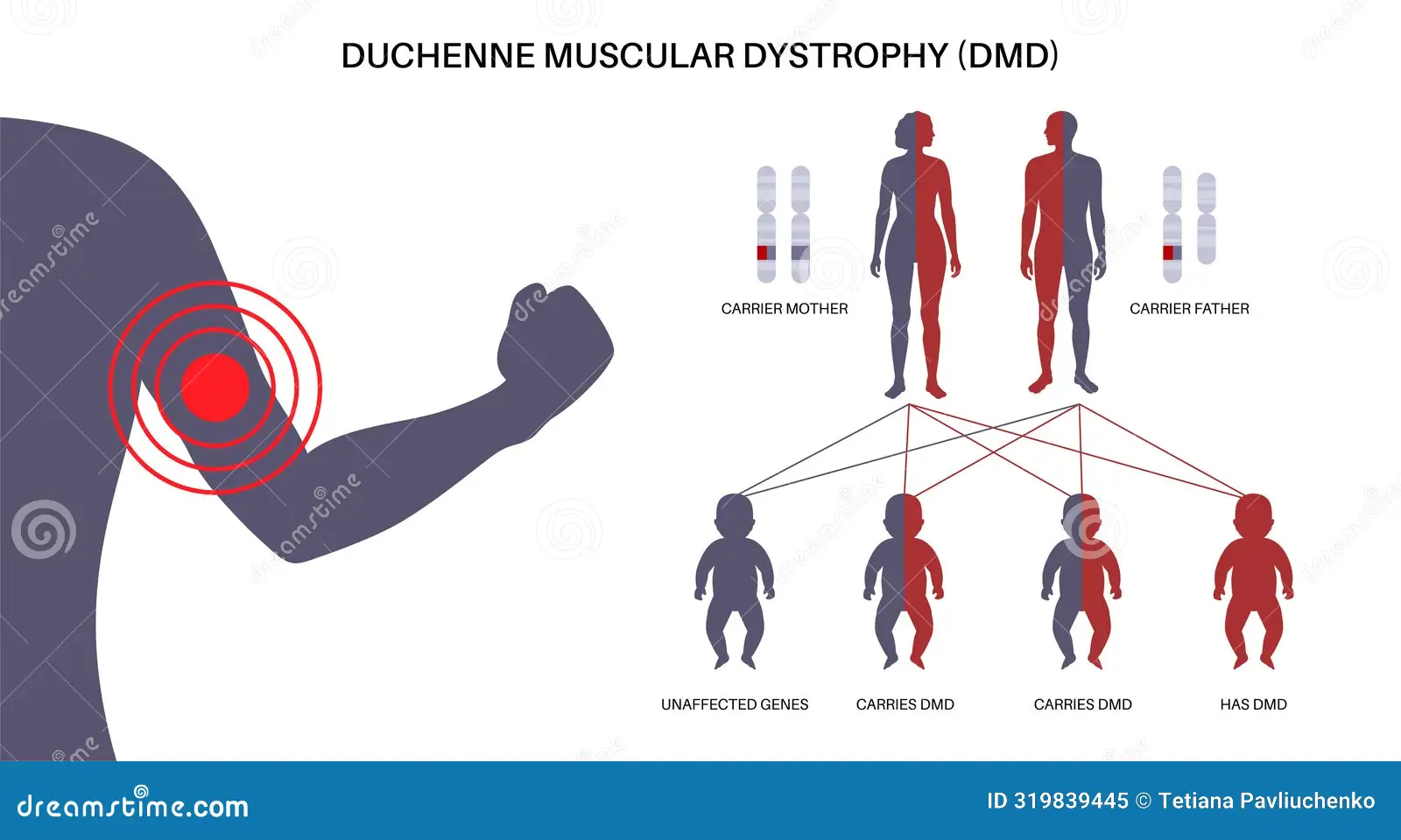
When 3-year-old Arjun started having difficulty climbing stairs and kept falling more frequently than other children his age, his pediatrician noticed enlarged calf muscles and ordered a blood test showing extremely elevated creatine kinase levels over 10,000 U/L (normal is less than 200), leading to genetic testing that confirmed Duchenne muscular dystrophy (DMD)—the most common fatal genetic disorder diagnosed in childhood, affecting approximately 1 in 3,500-5,000 male births worldwide, caused by mutations in the DMD gene on the X chromosome that prevent production of dystrophin, a crucial protein that protects muscle fibers during contraction. His neurologist explained that without dystrophin, every muscle movement causes tiny tears in muscle cell membranes that accumulate over time, leading to progressive muscle destruction and replacement with fat and scar tissue, resulting in a predictable devastating course where boys lose the ability to walk by ages 8-14, develop life-threatening heart and breathing problems by their teenage years, and historically died in their late teens or early twenties from cardiac or respiratory failure. However, the doctor also offered cautious hope: corticosteroid medications can slow disease progression by several years, multidisciplinary care focusing on cardiac and respiratory support has extended survival into the thirties and beyond, and revolutionary new therapies including exon-skipping drugs, gene therapy, and other emerging treatments are showing promise in clinical trials, transforming DMD from a rapidly fatal childhood disease to a chronic condition where many young men now survive into adulthood with good quality of life. Understanding Duchenne muscular dystrophy is crucial because early diagnosis (ideally by ages 3-5 before significant muscle damage accumulates) allows timely initiation of corticosteroids and supportive care that significantly improve outcomes, newborn screening for DMD is being implemented in some regions allowing even earlier intervention before symptoms appear, the disease follows a predictable progression requiring proactive management of cardiac complications (cardiomyopathy developing in nearly 100% of patients by late teens), respiratory muscle weakness (requiring nighttime ventilation then 24-hour support), orthopedic issues (scoliosis, contractures), and emerging therapies like eteplirsen, golodirsen, casimersen (exon-skipping drugs for specific mutations), and experimental gene therapies offer genuine hope for altering disease course, though they’re not cures and have significant limitations.
The Dystrophin Gene and Muscle Fiber Protection: When a Critical Protein Is Missing
Duchenne muscular dystrophy results from mutations in the DMD gene located on the X chromosome at position Xp21. The DMD gene is enormous—it’s the largest known human gene, spanning 2.4 million base pairs (about 0.1% of the entire human genome) and containing 79 exons (protein-coding segments). This massive size makes it particularly vulnerable to mutations. DMD provides instructions for making dystrophin, a large rod-shaped protein found in muscle cells. Dystrophin is part of a protein complex called the dystrophin-glycoprotein complex that connects the internal cytoskeleton of muscle fibers to the extracellular matrix outside the cell. This connection is critical because when muscles contract, enormous mechanical forces are generated. Dystrophin acts as a shock absorber, distributing these forces across the muscle cell membrane to prevent damage. In normal muscle, dystrophin makes up only about 0.002% of total muscle protein, yet this tiny amount is absolutely essential for muscle survival. Without dystrophin, every muscle contraction causes micro-tears in the cell membrane, allowing calcium to flood into the cell, triggering a cascade of damage including activation of enzymes that destroy muscle proteins, inflammation and immune response, and eventual muscle fiber death and replacement with fat and fibrous scar tissue.
The DMD gene mutations causing Duchenne are severe—they typically cause complete absence of functional dystrophin protein. About 60-70% of DMD cases result from large deletions (missing chunks of the DMD gene, typically involving one or more exons), 5-10% from large duplications (extra copies of gene segments), 25-30% from point mutations or small insertions/deletions (tiny changes that still destroy gene function), and rare cases from other complex rearrangements. Most mutations (around 70-80%) cause frameshift mutations—deletions or duplications that shift the reading frame, creating a premature stop codon and producing no functional protein. About 10-20% are nonsense mutations creating premature stop codons. The key is that dystrophin levels in Duchenne are less than 3% of normal (usually 0%), compared to Becker muscular dystrophy (a milder related condition) where dystrophin is 20-90% of normal—partially functional but abnormal. DMD follows X-linked recessive inheritance. Females have two X chromosomes—if one carries a DMD mutation, the other normal copy usually compensates, making them carriers but unaffected (though about 10% of female carriers have mild symptoms from skewed X-inactivation). Males have one X chromosome (plus one Y)—if their X carries a DMD mutation, they have no backup copy and develop Duchenne. About two-thirds of cases are inherited from carrier mothers (each pregnancy has 50% chance of passing mutated X to sons or daughters). About one-third are de novo mutations (new mutations occurring in the egg or early embryo—not inherited from parents). Carrier frequency in females is approximately 1 in 3,500, explaining the disease incidence in males.
Symptoms: A Predictable Devastating Progression Through Childhood and Adolescence
Duchenne muscular dystrophy follows a characteristic, heartbreakingly predictable progression. Early childhood (ages 2-5) shows subtle signs often missed initially including delayed motor milestones—walking later than peers (typically 15-18 months versus 12-15 months), difficulty running, jumping, climbing stairs, frequent falls and clumsiness—boys fall more than peers, struggle to keep up with activities, enlarged calf muscles (pseudohypertrophy)—calves appear muscular but it’s actually fat and scar tissue replacing muscle, Gower’s sign—difficulty standing from floor, using hands to “walk up” their own legs, and toe-walking—walking on toes because tight heel cords. Blood tests show massively elevated creatine kinase (CK)—10,000-30,000 U/L versus normal under 200 (CK is an enzyme released from damaged muscles). Middle childhood (ages 5-10) brings progressive proximal weakness (shoulders, hips, thighs weaken first)—difficulty climbing stairs (needing handrail, going one step at a time), difficulty getting up from sitting or lying, waddling gait from hip muscle weakness, and frequent falls. Muscle contractures begin developing as muscles tighten, particularly heel cords (Achilles tendons) causing toe-walking. Fatigue increases with decreased stamina and endurance. Some boys develop learning difficulties—about 30% have cognitive impairments or learning disabilities (dystrophin is also expressed in brain, though brain problems don’t progress like muscle problems).
Early adolescence (ages 8-14) marks loss of walking ability—most boys transition to wheelchair between ages 9-12 (earlier without corticosteroids, later with treatment), progressive upper extremity weakness making self-feeding, writing, using computer difficult, scoliosis (spinal curvature) developing rapidly once wheelchair-bound in 90%+ of boys, affecting sitting balance and compressing lungs, and contractures worsening in elbows, knees, ankles despite stretching. Respiratory muscle weakness begins—measured by declining vital capacity (lung volume), initially asymptomatic but progressive. Cardiac involvement starts—dilated cardiomyopathy (heart muscle weakening and enlarging) beginning in early teens though often asymptomatic initially. Late adolescence and young adulthood (ages 15+) show severe disability with complete wheelchair dependence, severe upper extremity weakness—many lose ability to feed themselves, use phones, control wheelchair independently, respiratory failure developing—nighttime hypoventilation first (CO2 retention during sleep), then daytime respiratory insufficiency, requiring non-invasive ventilation (BiPAP) initially at night, progressing to 24-hour ventilator dependence. Progressive cardiomyopathy with nearly 100% of patients developing heart muscle disease by late teens, some developing heart failure symptoms (fatigue, shortness of breath, swelling), and ongoing orthopedic complications including severe scoliosis (often requiring surgery), complete loss of independent mobility, and dysphagia (swallowing difficulties) in some patients.
Historically (before modern care), most boys died by late teens or early twenties from respiratory failure (most common, especially overnight during sleep—respiratory muscles too weak to breathe adequately), cardiac failure or arrhythmias (second most common cause), or complications from severe disability (infections, aspiration pneumonia). With modern multidisciplinary care (corticosteroids, cardiac medications, ventilation support), median survival has extended significantly to early-mid thirties, with many patients living into their forties or even fifties with optimal care. Quality of life varies enormously depending on access to care, ventilation support, social support, and individual factors.
Diagnosis: From Clinical Suspicion to Genetic Confirmation
Diagnosing DMD requires recognizing the clinical pattern and confirming with specific testing. Clinical suspicion arises from delayed motor milestones or difficulty with gross motor skills in young boy, frequent falls, difficulty climbing stairs, running in toddler/preschooler, enlarged calf muscles plus muscle weakness, Gower’s sign (using hands to stand from floor), family history of DMD or unexplained male infant/child deaths, and elevated CK on routine blood work (sometimes found incidentally). Initial laboratory testing shows massively elevated creatine kinase (CK)—typically 10,000-30,000 U/L or higher versus normal under 200. CK is very sensitive for DMD, with levels elevated from birth (even before symptoms). However, CK is non-specific (elevated in many muscle conditions), so genetic testing is needed for confirmation. Genetic testing provides definitive diagnosis using DMD gene deletion/duplication testing (MLPA—multiplex ligation-dependent probe amplification or chromosomal microarray) detecting large deletions or duplications—identifies mutation in about 70% of DMD patients. If negative, DMD gene sequencing (full gene sequencing) identifies point mutations, small insertions/deletions—finds remaining 30% of mutations. Together, these tests diagnose greater than 99% of DMD cases.
Muscle biopsy (rarely needed now) was historically used showing complete or near-complete absence of dystrophin on immunostaining (less than 3% of normal, usually 0%), muscle fiber degeneration and regeneration, fat and fibrous tissue replacing muscle, and inflammatory infiltrates. Biopsy is now rarely performed since genetic testing is definitive, less invasive, and provides more information (specific mutation guides potential treatment eligibility for exon-skipping drugs). Additional testing after diagnosis includes cardiac evaluation with echocardiogram establishing baseline heart function and EKG detecting early cardiac abnormalities. Pulmonary function tests establish baseline lung function (vital capacity, maximum inspiratory/expiratory pressures). Developmental and cognitive assessment screens for learning disabilities. Genetic counseling provides family education, carrier testing for female relatives, and reproductive counseling. Newborn screening for DMD is being piloted or implemented in some regions (New York State, some European countries) using CK screening or genetic testing, allowing presymptomatic diagnosis and earlier intervention. However, it remains controversial due to lack of curative treatment and psychological impact of diagnosis.
Treatment: Corticosteroids, Multidisciplinary Care, and Emerging Therapies
While DMD has no cure, treatment has improved dramatically. Corticosteroid therapy is the current standard of care using prednisone or deflazacort (a corticosteroid with potentially fewer side effects) starting around ages 4-6, before significant functional decline. Daily dosing (0.75 mg/kg prednisone or 0.9 mg/kg deflazacort) is standard, with weekend-only dosing showing less efficacy. Benefits include prolonging walking ability by 2-3 years on average (boys walking to age 12-14 versus 9-11 without treatment), slowing respiratory muscle decline (delaying need for ventilation), slowing cardiac disease progression, reducing scoliosis severity, and improving overall survival. Side effects include weight gain and obesity (nearly universal), growth suppression and short stature, bone loss and fractures (osteoporosis from steroids plus immobility), behavioral changes (mood swings, emotional lability), cushingoid features (moon face, buffalo hump), increased infection risk, cataracts (rare), and glucose intolerance. Side effect management requires calcium and vitamin D supplementation, bisphosphonates for bone health, dietary management to control weight, behavioral support, and monitoring for complications. Despite side effects, benefits strongly outweigh risks—corticosteroids are the only medication proven to slow DMD progression across multiple outcomes.
Cardiac management is critical with ACE inhibitors or ARBs starting by age 10-12 (even before cardiac symptoms) to prevent or delay cardiomyopathy. Beta-blockers are added if needed. Regular echocardiograms monitor cardiac function every 1-2 years initially, more frequently if abnormalities develop. Advanced heart failure may require more intensive medications, devices (pacemakers, defibrillators), or rarely, heart transplant. Respiratory care includes pulmonary function testing every 6-12 months tracking vital capacity decline, cough assist devices helping clear secretions when cough becomes weak, and non-invasive ventilation (BiPAP) starting when vital capacity drops below 50% predicted or nocturnal hypoventilation develops. Initially needed only at night, this progresses to 24-hour ventilation in most patients. Tracheostomy and invasive ventilation is used for 24-hour support in some patients, though many use non-invasive ventilation via mask instead. Routine vaccinations including annual influenza vaccine and pneumococcal vaccine are important as respiratory infections are major causes of complications.
Physical therapy and orthopedic management involve stretching and range of motion exercises preventing contractures, ankle-foot orthoses (AFOs) maintaining ankle position and prolonging walking, knee-ankle-foot orthoses (KAFOs) for standing/walking support in some patients, and scoliosis management with bracing (often ineffective in DMD) or spinal fusion surgery for severe progressive curves. Surgery is controversial given surgical risks but can improve sitting balance and slow respiratory decline. Nutritional support addresses obesity from steroids and inactivity through dietary counseling, and dysphagia and feeding difficulties in advanced disease, sometimes requiring feeding tubes.
Emerging disease-modifying therapies show promise. Exon-skipping drugs (eteplirsen, golodirsen, casimersen, viltolarsen) are FDA-approved antisense oligonucleotides that cause the cellular machinery to skip specific exons during dystrophin mRNA processing, restoring the reading frame and allowing production of shortened but partially functional dystrophin (converting Duchenne-like pattern to Becker-like). These are mutation-specific—only work for patients with deletions amenable to skipping specific exons (about 13% amenable to exon 51 skipping—eteplirsen, 8% to exon 53—golodirsen, 8% to exon 45—casimersen). Given IV every 1-2 weeks for life, they have modest efficacy (producing 1-15% of normal dystrophin levels, slowing but not halting progression) with controversy about clinical benefit (FDA accelerated approval based on dystrophin production, but clinical benefit data are mixed). Ataluren targets nonsense mutations (10-15% of DMD patients) helping cellular machinery read through premature stop codons. Oral medication three times daily shows modest benefit in slowing progression. Approved in Europe but not US.
Gene therapy using micro-dystrophin (shortened dystrophin gene delivered via AAV vector in single IV infusion) is in clinical trials showing dystrophin production in muscle biopsies and possible functional stabilization, though long-term efficacy and safety unknown. Multiple trials ongoing including SRP-9001 (ongoing Phase 3 trials). Other approaches being studied include myostatin inhibition (blocking myostatin protein that limits muscle growth), utrophin upregulation (increasing expression of utrophin, a protein similar to dystrophin), and CRISPR gene editing (experimental—correcting the mutation directly in muscle cells). These are years away from approval.
Living with DMD: Progressive Disability, Family Impact, and Quality of Life
Living with Duchenne muscular dystrophy means adapting to progressive physical limitations while maintaining quality of life and hope. The reality for boys and families involves early childhood often appearing relatively normal with boys participating in preschool, playing with peers (though unable to keep up physically), and early interventions (PT, OT, speech therapy if needed) starting. Middle childhood brings increasing limitations—transitioning to special education with accommodations, using assistive technology (computers replacing handwriting), and developing adaptive strategies. Adolescence marks major transitions—loss of walking (emotionally devastating for boys and families), wheelchair dependence (power wheelchair for independence), onset of visible disability affecting social interactions and self-image, and navigating school with increasing physical limitations. Young adulthood shows complete care dependence requiring assistance with all activities of daily living (bathing, dressing, toileting, eating), 24-hour ventilator dependence in many, decisions about tracheostomy versus non-invasive ventilation, and transition to adult medical care and living arrangements (many live with family, some in group homes or assisted living).
Psychological and social impact is profound with normal intelligence meaning full awareness of disease and prognosis, anxiety and depression common in patients and families, social isolation from progressive disability, relationship challenges (many young men with DMD desire romantic relationships but face physical and social barriers), and grief over lost abilities and shortened lifespan. However, quality of life can be good with modern technology providing independence (power wheelchairs, computers, communication devices), education and career possibilities (many complete high school, some attend college online or adapted programs, work in computer-based careers), strong social connections and community (DMD family networks, online communities), hobbies and interests adapted to abilities, and meaning and purpose despite physical limitations.
Family impact is enormous including 24/7 caregiving burden (exhausting for parents, particularly mothers who often become primary caregivers), financial stress (equipment, modifications, medical care, potential loss of parental income), sibling impact (siblings may feel neglected, assume caregiving roles, carry emotional burden), and marital stress (many marriages don’t survive the strain of caring for child with DMD). Support resources include the Muscular Dystrophy Association providing clinics, equipment, support groups, and research funding. Parent Project Muscular Dystrophy focuses specifically on DMD advocacy and research. CureDuchenne funds research toward treatments and cure. Many smaller foundations and support groups offer assistance.
The future holds hope with gene therapy potentially offering major benefit within 5-10 years, exon-skipping and other molecular therapies continually improving, multidisciplinary care extending survival and quality of life, and the possibility that boys diagnosed today may live significantly longer with better function than previous generations. While DMD remains a devastating disease, it’s no longer the rapidly fatal childhood condition it once was—it’s now a serious chronic disease where many young men survive into their thirties, forties, or beyond, maintaining relationships, pursuing education and careers, and living meaningful lives despite profound physical limitations.
Frequently Asked Questions
Q1: At what age do boys with Duchenne muscular dystrophy typically stop walking, and can anything delay this?
Most boys with Duchenne stop walking between ages 9-12, though the range varies from as early as 7 to as late as 16 depending on several factors. The single most important factor affecting walking duration is corticosteroid treatment—boys treated with daily prednisone or deflazacort typically walk until age 12-14 (some even to 15-16), while untreated boys typically lose walking by age 9-11, representing a 2-3 year extension of walking ability. Physical therapy with stretching and range of motion exercises helps maintain flexibility and delay contractures that interfere with walking, while ankle-foot orthoses (AFOs—leg braces) maintain ankle position and can prolong walking by months to a year. Maintaining healthy weight is crucial since obesity from steroids makes walking harder—dietary management is important. Individual genetic factors matter as some DMD mutations cause slightly slower progression than others, though all eventually cause walking loss. Early intervention starting corticosteroids by ages 4-6 (before significant decline) provides maximum benefit. Once walking is lost, it’s essentially never regained—progressive muscle loss is irreversible, making prevention (delaying loss through treatment) critical.
Q2: What are the main side effects of corticosteroids in DMD, and are they worth the risks?
Corticosteroids (prednisone, deflazacort) cause significant side effects that families and doctors must carefully manage, yet the overwhelming consensus is that benefits far outweigh risks for most boys with DMD. The most common and troublesome side effects include weight gain and obesity (nearly universal—boys on steroids gain significant weight, which can make mobility harder and increase cardiac/respiratory strain), short stature from growth suppression (boys end up several inches shorter than they would without steroids), bone problems including osteoporosis and increased fracture risk from both steroids and immobility (vertebral compression fractures are common), behavioral changes such as mood swings, emotional lability, irritability (some boys become difficult to manage behaviorally), cushingoid appearance with moon face, buffalo hump, and stretch marks, and increased infection susceptibility (though serious infections are uncommon). Less common but serious side effects include cataracts (especially with long-term use—regular eye exams needed), glucose intolerance or diabetes (monitoring blood sugar needed), and gastrointestinal issues like gastritis or ulcers. However, the benefits are profound and well-documented—prolonging walking by 2-3 years (maintaining independence and development), slowing respiratory decline by years (delaying ventilator need), slowing cardiac disease (reducing heart failure and improving survival), reducing scoliosis severity (preventing or delaying spinal surgery), and improving overall survival by roughly 5-10 years. Every major DMD clinical guideline strongly recommends corticosteroids as standard of care starting around ages 4-6. The key is managing side effects proactively through calcium and vitamin D supplementation plus bisphosphonates for bone health, careful dietary management to control weight gain, behavioral support and sometimes medications for mood issues, and regular monitoring for complications. Some families choose to stop steroids due to intolerable side effects, but this should be discussed carefully with the DMD care team given the significant benefits being lost.
Q3: My son was just diagnosed with Duchenne at age 4. What should we do immediately, and what does his future look like?
First, take a breath—you’re not alone, and while DMD is a serious progressive disease, modern care has transformed outcomes significantly. Immediate actions in the first weeks to months include meeting with a neuromuscular specialist at an MDA clinic or comprehensive DMD center (these centers have multidisciplinary teams experienced in DMD care), starting corticosteroids (prednisone or deflazacort) if your son is age 4+ and not yet on them—this is the single most important treatment, establishing baseline assessments with cardiac echo, pulmonary function tests (if your son can cooperate—may need to wait until age 5-6), and developmental/cognitive evaluation. Beginning physical therapy focusing on stretching and range of motion is essential, as is connecting with support organizations (MDA, Parent Project Muscular Dystrophy, local DMD family groups for emotional support and practical advice), genetic counseling for family planning and testing of female relatives for carrier status, and exploring clinical trials (some trials enroll newly diagnosed boys—ask your neurologist). His likely future trajectory at ages 4-8 involves appearing relatively normal—attending regular school (with accommodations), playing with peers (though can’t keep up physically), starting to show obvious weakness (difficulty stairs, frequent falls), and remaining generally independent in most activities. Ages 8-14 bring progressive weakness, loss of walking ability (around age 10-14 with steroids), transition to wheelchair, and upper extremity weakness developing. Teenage years show complete wheelchair dependence, progressive upper extremity weakness, scoliosis developing, need for nighttime ventilation starting (typically ages 15-18), and cardiac involvement developing. Young adulthood involves 24-hour ventilator dependence likely, complete care dependence for all ADLs, ongoing cardiac management, and need for long-term care planning. However, this trajectory is variable and the outlook is improving—boys diagnosed and treated today are expected to live into their thirties, forties, or beyond (versus teens-twenties historically). Quality of life can be good despite severe physical limitations—normal intelligence allows education, relationships, work (computer-based), hobbies, and meaningful life. Critical things to know include early treatment (steroids, proactive cardiac/respiratory care) dramatically improves outcomes, preparing for transitions (loss of walking, ventilation needs) emotionally and practically helps families cope, staying connected to the DMD community provides support and information, maintaining hope while being realistic about progression allows families to live fully while planning appropriately, and research is advancing rapidly with gene therapies and other treatments in trials potentially benefiting your son. This is not the death sentence it once was—it’s now a serious chronic disease requiring intensive management but compatible with decades of life.
Q4: Are there any new treatments or gene therapies that could cure or significantly improve Duchenne?
The DMD research landscape is very active with multiple promising approaches in various stages of development, though it’s important to balance hope with realistic expectations about what’s currently available and what’s still experimental. Currently available treatments include exon-skipping drugs (eteplirsen, golodirsen, casimersen, viltolarsen—FDA approved) which are antisense oligonucleotides that restore some dystrophin production by skipping specific exons. They’re mutation-specific—only about 30% of DMD patients have deletions amenable to currently available exon-skipping drugs. Efficacy is modest—they produce 1-15% of normal dystrophin levels (better than 0% but far less than the 20-30% needed for Becker-like mild disease). Clinical benefit is debated—some studies show slowing of progression, others show minimal functional benefit. They require IV infusions every 1-2 weeks for life and cost $300,000+ annually. Ataluren (Translarna) targets nonsense mutations (10-15% of patients), helping cells read through premature stop codons. Approved in Europe but not the US. Oral medication showing modest benefit in slowing progression. Corticosteroids remain the most effective currently available treatment despite being non-specific to DMD. Experimental therapies in clinical trials include gene therapy with micro-dystrophin using AAV vectors delivering a shortened dystrophin gene via single IV infusion. Multiple trials ongoing including SRP-9001 (Sarepta) in Phase 3 trials with mixed results so far—some patients showing dystrophin production and possible functional stabilization, others showing minimal benefit, and concerns about durability (does one dose last for years or decades?). Results expected in next 1-2 years will determine if this approaches FDA approval. CRISPR gene editing is attempting to directly correct the DMD mutation in muscle cells and is in very early trials (Phase 1/2) with preliminary data showing safety but limited efficacy data so far. This is years from approval even if successful. Utrophin upregulation tries to increase expression of utrophin (a protein similar to dystrophin) to compensate for missing dystrophin, and is in clinical trials with mixed early results. Realistic timeline and expectations show exon-skipping drugs are available now but have limited applicability (only certain mutations) and modest benefit. Gene therapy may be approved in 2-5 years if current trials succeed, potentially offering superior benefit to exon-skipping but still not a cure (unlikely to restore normal strength—more likely to slow progression). CRISPR and other approaches are 10+ years away at minimum given they’re still in early-phase trials. Nothing currently available or in near-term pipeline is curative—at best, these treatments may convert Duchenne-like progression to something closer to Becker muscular dystrophy (slower progression, longer lifespan, better function). Your son should absolutely be evaluated for clinical trial eligibility—trials advance the field and may provide access to experimental treatments. However, maintain realistic expectations and continue standard care (corticosteroids, cardiac/respiratory management) which remain the foundation of DMD treatment even as new therapies emerge.
Q5: I’m a carrier of a DMD mutation. What are the chances my children will have Duchenne, and what are my options?
As a confirmed DMD carrier (one normal DMD gene, one mutated DMD gene on your X chromosomes), the inheritance pattern depends on the sex of each child. For sons, each pregnancy has a 50% (1 in 2) chance he inherits your normal X chromosome and is unaffected, and 50% chance he inherits your mutated X chromosome and has Duchenne muscular dystrophy. For daughters, each pregnancy has 50% chance she inherits your normal X chromosome (she’ll be unaffected, non-carrier), and 50% chance she inherits your mutated X chromosome (she’ll be a carrier like you but almost certainly unaffected clinically, though 10% of female carriers have mild symptoms). These probabilities apply independently to each pregnancy. Your reproductive options include accepting the 50% risk for sons and relying on prenatal diagnosis during each pregnancy—chorionic villus sampling (CVS) at 10-13 weeks or amniocentesis at 15-20 weeks determines if fetus is male and if so, whether he has the DMD mutation. If affected male fetus, some families choose termination while others continue pregnancy with knowledge allowing preparation. If female fetus or unaffected male, pregnancy continues with reassurance. Small miscarriage risk (0.1-0.5%) from testing. Preimplantation genetic diagnosis (PGD) with IVF creates embryos via IVF, tests each embryo for the DMD mutation, and transfers only unaffected or carrier female embryos (avoiding affected males). This ensures no affected children. Advantages include eliminating DMD risk and avoiding prenatal testing/termination decisions. Disadvantages include very expensive ($15,000-30,000+ per cycle), physically demanding IVF process, no pregnancy guarantee, and ethical concerns for some families. Using donor eggs from a non-carrier eliminates DMD risk entirely (all children would have normal DMD genes), though involves genetic material outside your relationship. Adoption avoids genetic risks with different challenges. Not having biological children is a personal choice some make. Factors to consider include your family history—have you had affected relatives? What was their experience? This may influence your risk tolerance. What are your personal values and beliefs about prenatal testing, termination, or embryo selection? These vary greatly and guide acceptable options. What are your financial circumstances? PGD is expensive. What’s your emotional capacity for managing DMD in a child if it occurs? There’s no objectively “right” choice—families make different decisions based on their values, circumstances, and risk tolerance. My recommendation is to meet with a genetic counselor who can discuss your specific mutation, explain all options thoroughly, help you think through what’s right for your family, and potentially connect you with other DMD carrier families who’ve faced these decisions. Whatever you choose, support is available through DMD organizations, genetic counselors, and family support groups to help you navigate this decision and whatever path you take.
Disclaimer
This article adapts publicly available information from medical databases and research organizations. This content is for informational and educational purposes only and does not constitute medical advice. ObserverVoice.com is a news and information platform — not a healthcare provider. Decisions about Duchenne muscular dystrophy diagnosis, genetic testing, corticosteroid treatment, cardiac and respiratory management, and emerging therapies should be made in consultation with qualified physicians, pediatric neurologists, neuromuscular specialists, cardiologists, pulmonologists, geneticists, and multidisciplinary DMD care teams who can evaluate your individual situation, genetic mutations, cardiac and respiratory function, and health circumstances. If you have questions about DMD diagnosis or treatment options, please consult with your neuromuscular team.
References
- Muscular Dystrophy Association. Duchenne Muscular Dystrophy (DMD). https://www.mda.org/disease/duchenne-muscular-dystrophy
- Parent Project Muscular Dystrophy. About Duchenne. https://www.parentprojectmd.org/about-duchenne/
- PMC. Duchenne Muscular Dystrophy: Current Therapeutic Approaches. https://www.ncbi.nlm.nih.gov/pmc/articles/PMC8308746/
- PMC. Duchenne Muscular Dystrophy: From Diagnosis to Treatment. https://www.ncbi.nlm.nih.gov/pmc/articles/PMC7460519/
- World Health Organization. Genomic Resource Centre. https://www.who.int/teams/genomics-and-digital-health
Observer Voice is the one stop site for National, International news, Sports, Editor’s Choice, Art/culture contents, Quotes and much more. We also cover historical contents. Historical contents includes World History, Indian History, and what happened today. The website also covers Entertainment across the India and World.

