

Celiac Disease: Beyond Gluten Sensitivity — the Serious Autoimmune Reality

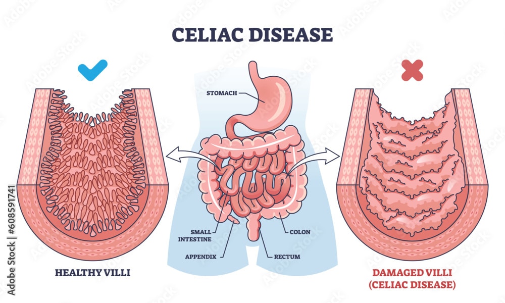
Imagine eating a food that triggers your immune system to attack your own intestines. Tiny fingerlike projections in your small intestine—villi—gradually flatten and disappear. Your intestines lose the ability to absorb nutrients. You become malnourished despite eating adequate food. You develop anemia, osteoporosis, and infertility. Your risk of intestinal cancer increases. This is celiac disease—a serious autoimmune disorder where the immune system reacts to gluten in wheat, barley, and rye, causing progressive destruction of the small intestine and systemic complications affecting multiple organs. Celiac disease is an autoimmune condition where the body produces antibodies against tissue transglutaminase (tTG)—an enzyme in the intestinal lining. When gluten is consumed, the immune system mistakenly identifies it as a threat. Antibodies and T cells attack the small intestine. The intestinal lining becomes inflamed. The villi—projections that increase absorptive surface area—gradually flatten. The intestines lose the ability to absorb nutrients. Malabsorption develops. Nutrient deficiencies occur. Symptoms develop from both intestinal damage and systemic nutrient deficiency. Celiac disease affects approximately 1 percent of the population worldwide. The disease is more common than previously recognized. Increased screening has improved diagnosis. However, many cases remain undiagnosed. The disease predominantly affects those of European ancestry. It is less common in Asian and African populations. What makes celiac disease particularly important is the distinction from non-celiac gluten sensitivity. Many people report gluten sensitivity without having celiac disease. Celiac disease is a specific autoimmune disease with intestinal damage and systemic complications. Non-celiac gluten sensitivity is a separate condition. True celiac disease is serious and requires lifelong gluten avoidance. With appropriate gluten-free diet, intestinal healing occurs. Nutrient absorption normalizes. Systemic complications can be prevented. In this comprehensive article, we will explore what celiac disease is, understand how gluten triggers autoimmune damage, recognize diverse symptoms beyond digestive complaints, learn about serious systemic complications, understand diagnostic methods, explore the gluten-free diet, and discover management strategies for achieving intestinal healing and preventing long-term complications.
Understanding Normal Intestinal Structure and Function
Before we explore celiac disease, we need to understand normal intestinal structure and nutrient absorption. The small intestine is the primary site of nutrient absorption. The small intestine consists of three portions: duodenum, jejunum, and ileum. The duodenum is the first 25 centimeters—receives partially digested food from the stomach. The jejunum is the middle 2.5 meters—primary site of nutrient absorption. The ileum is the final 3.5 meters—absorbs remaining nutrients and vitamin B12. The intestinal lining consists of a single layer of epithelial cells. These cells form a barrier between intestinal contents and blood. The intestinal lining has specialized projections called villi. Villi are fingerlike projections extending into the intestinal lumen. Villi significantly increase absorptive surface area. Between villi are crypts where new epithelial cells are produced. New epithelial cells continuously replace old ones—the intestinal lining completely renews every 3 to 5 days. Epithelial cells are connected by tight junctions. These tight junctions form a barrier preventing bacteria and large molecules from passing. The intestinal barrier is selectively permeable. Water, electrolytes, and nutrients pass through. Bacteria and large molecules are excluded. Nutrient absorption occurs through specialized transporters. Glucose, amino acids, and fats are actively transported. Vitamins and minerals are absorbed by various mechanisms. The intestinal lining also contains immune cells. Gut-associated lymphoid tissue (GALT) lines the intestine. T cells and B cells recognize food antigens. Normally, immune tolerance develops to harmless food proteins. Immune response is suppressed to food. However, in celiac disease, immune response to gluten develops. The immune tolerance to gluten fails. Antibodies against gluten are produced. T cells attack gluten-containing cells. The immune attack damages intestinal villi. The villi flatten. The absorptive surface area decreases. Nutrient absorption decreases. Malabsorption develops. Understanding normal intestinal structure helps explain how intestinal damage causes nutrient deficiency.
What is Celiac Disease?
Celiac disease is an autoimmune disorder triggered by gluten consumption. Gluten is a protein found in wheat, barley, and rye. In susceptible individuals, gluten triggers immune response. The immune system produces antibodies against tissue transglutaminase (tTG). The autoantibodies bind to tTG in the intestinal lining. The antibody-antigen complexes are recognized as foreign. Complement is activated. Inflammatory cells infiltrate intestinal villi. CD8+ cytotoxic T cells destroy intestinal epithelial cells. Th1 cells produce inflammatory cytokines. The inflammatory environment damages villi. The villous epithelium is destroyed. The villi progressively flatten. The villous atrophy is characteristic of celiac disease. As villi flatten, absorptive surface area decreases. Nutrient absorption becomes impaired. Malabsorption develops. Iron absorption decreases. Calcium absorption decreases. Fat-soluble vitamin absorption decreases. Protein absorption decreases. Eventually, severe malabsorption develops. What causes immune response to gluten in celiac disease is incompletely understood. Genetic factors are important. HLA-DQ2 and HLA-DQ8 genes increase susceptibility. Approximately 90 to 95 percent of celiac disease patients have HLA-DQ2. The remaining 5 to 10 percent have HLA-DQ8. However, these HLA types are found in approximately 30 to 40 percent of the general population. Most people with HLA-DQ2/DQ8 never develop celiac disease. Gluten exposure is necessary. Without gluten consumption, celiac disease does not develop. Introducing gluten in infancy (between 4 and 12 months) increases celiac disease risk. Environmental factors trigger disease. Infections have been suspected. Rotavirus and adenovirus have preceded celiac disease development. The infections might trigger immune response cross-reacting against gluten. Intestinal dysbiosis—abnormal bacterial composition—might contribute. Specific bacteria promote immune tolerance. Dysbiosis might reduce this protection. Stress might trigger disease. Psychological stress preceding celiac disease development has been reported. Stress might activate the immune system. The combination of genetic predisposition, gluten exposure, and environmental trigger results in celiac disease development. Celiac disease is classified into two main presentations. Classical celiac disease involves gastrointestinal symptoms. Non-classical celiac disease involves minimal or no gastrointestinal symptoms. Non-classical disease often causes systemic symptoms. Diagnosis is delayed in non-classical presentation. Latent celiac disease is serology-positive (antibodies present) but with normal intestinal biopsy. Potential celiac disease is serology-positive but without villous atrophy yet. These early stages might progress to overt celiac disease if untreated.
Recognizing Symptoms: Beyond Digestive Complaints
Celiac disease symptoms are highly variable. Some patients have only gastrointestinal symptoms. Others have systemic symptoms dominating the presentation. Recognizing the diversity of symptoms helps diagnosis. Diarrhea is the most common gastrointestinal symptom. Stools become loose and frequent. Diarrhea develops after eating gluten. Some patients have diarrhea occurring hours after exposure. Steatorrhea—fatty stools that float—develops from fat malabsorption. The stools are pale, bulky, and foul-smelling. Abdominal pain develops. Cramping pain occurs. Pain is often postprandial—after eating. Bloating develops. Abdominal distension from gas occurs. Patients report feeling uncomfortably full after small meals. Constipation develops in some patients—contrasting with diarrhea in others. Abdominal symptoms vary. Nausea develops. Vomiting occurs in some patients. Loss of appetite develops. Weight loss occurs from malabsorption. Despite eating adequately, weight gradually decreases. In children, failure to thrive develops—children do not gain weight normally. Growth restriction develops. Children remain shorter than expected. Anemia develops from iron malabsorption. Fatigue results from anemia. Weakness develops. Shortness of breath develops with exertion. Pallor—pale skin—indicates anemia. Bone pain develops from calcium deficiency. Osteoporosis—weak bones—develops from vitamin D and calcium malabsorption. Bones fracture easily. Height loss occurs from vertebral fractures. Dental problems develop. Enamel defects appear. Tooth decay becomes frequent. Delayed tooth eruption occurs in children. Dermatitis herpetiformis develops in some patients. This is an intensely itchy rash. Blisters form on elbows, knees, buttocks. The rash is pathognomonic for celiac disease. Aphthous ulcers develop—painful sores in mouth. Hair loss (alopecia) develops. Hair becomes thin and brittle. Hair growth is impaired. Joint and muscle pain develop. Muscle weakness develops from protein deficiency. Joint pain—arthralgia—occurs. Arthritis sometimes develops. Infertility develops. Women have difficulty conceiving. Miscarriage risk increases. Men have decreased sperm count. Decreased fertility results from nutrient deficiency and immune dysfunction. Menstrual irregularities develop. Periods become irregular or stop. Delayed menarche—first menstruation—occurs. Early menopause can develop. Neurological symptoms develop in some patients. Peripheral neuropathy—nerve damage—causes tingling and numbness. Ataxia—loss of coordination—develops. Headaches develop. Migraine becomes more frequent. Mood changes develop. Depression develops from nutrient deficiency. Anxiety develops. Irritability develops. Cognitive changes develop. Memory impairment develops. Concentration difficulty develops. Brain fog—difficulty thinking clearly—occurs. Seizures rarely develop from severe magnesium or calcium deficiency. Many celiac disease patients are asymptomatic. The disease is discovered on screening. Family members of diagnosed patients are screened. Screening detects disease before symptoms develop. Some patients have minimal symptoms attributed to other causes. Diagnosis delay is common—average delay is 6 to 10 years from symptom onset to diagnosis.
Understanding Progressive Intestinal Damage: Villous Atrophy and Beyond
Understanding how gluten-triggered immune attack progressively damages intestines helps explain disease progression. The immune attack is triggered by gluten consumption. The intestinal immune system recognizes gluten peptides. Dendritic cells present gluten peptides to CD4+ T cells. CD4+ T cells become activated. The activated T cells produce inflammatory cytokines. TNF-alpha and interferon-gamma are produced. The inflammatory cytokines activate CD8+ cytotoxic T cells. CD8+ T cells directly kill intestinal epithelial cells. The killed epithelial cells are replaced by immune cells. Additionally, B cells produce antibodies against tissue transglutaminase. Antibody-tTG complexes form. These complexes activate complement. Complement-mediated cell lysis damages epithelial cells. The epithelial damage is progressive. Initially, intraepithelial lymphocytes increase. The lymphocytes infiltrate between epithelial cells. Crypt hyperplasia develops—crypts enlarge. Villous height decreases—villi become shorter. The villous-to-crypt ratio decreases. Progressive villous atrophy develops. The villi become increasingly flattened. Eventually, complete villous atrophy develops—villi are completely absent. The intestinal lining becomes flat. This complete villous atrophy is the most severe form. The progression from normal villi to complete atrophy occurs over months to years. The rate of progression varies. Some patients develop severe atrophy rapidly. Others develop it gradually. Intestinal permeability increases. The damaged epithelium has leaky tight junctions. Bacteria and bacterial lipopolysaccharides (LPS) cross the barrier. The bacterial antigens trigger immune activation. Systemic inflammation develops. The increased permeability allows larger food molecules to cross. The immune system produces antibodies against these food molecules. Multiple food sensitivities can develop. As intestinal damage progresses, nutrient absorption decreases. Iron absorption decreases—anemia develops. Calcium absorption decreases—osteoporosis develops. Fat-soluble vitamins (A, D, E, K) absorption decreases. Vitamin A deficiency causes night blindness. Vitamin D deficiency causes bone disease. Vitamin E deficiency causes neuropathy. Vitamin K deficiency causes bleeding tendency. Fat absorption is severely impaired. Steatorrhea develops. Fat-soluble vitamin deficiency results. Protein absorption decreases. Protein malnutrition develops. Albumin production decreases. Edema develops from low albumin. Immune function suffers from protein deficiency. Lactose intolerance develops. The damaged intestine produces less lactase enzyme. Cow’s milk becomes poorly tolerated. Lactose malabsorption causes bloating and diarrhea. The intestinal damage and nutrient deficiency cause systemic complications.
Systemic Complications: Beyond the Intestines
Untreated celiac disease causes serious complications affecting multiple organ systems. Understanding these complications emphasizes the importance of early diagnosis and treatment. Anemia is extremely common. Iron deficiency anemia develops from iron malabsorption. Hemoglobin decreases. Oxygen-carrying capacity decreases. Fatigue and shortness of breath result. Macrocytic anemia develops from vitamin B12 or folate deficiency. Large red blood cells indicate deficiency. Fatigue and neurological symptoms result. Hemolytic anemia rarely develops. Autoimmune destruction of red blood cells occurs. Osteoporosis develops from calcium and vitamin D deficiency. Bone mineral density decreases. Bones become fragile. Fractures occur from minor trauma. Height loss occurs from vertebral fractures. Osteoporosis increases with advancing age. Young patients with untreated celiac disease already have low bone density. Thyroid disease develops frequently. Hashimoto’s thyroiditis develops in approximately 5 percent of celiac disease patients. Hypothyroidism results. Fatigue and weight gain worsen. Graves’ disease develops in some. Hyperthyroidism results. The increased frequency results from molecular mimicry—thyroid peroxidase antigens resemble gluten-derived peptides. Neurological complications develop. Peripheral neuropathy causes numbness and tingling. Ataxia causes loss of coordination and balance. Seizures develop from electrolyte deficiency. Migraine becomes more frequent. Neurological complications result from nutrient deficiency and autoimmunity. Infertility and miscarriage. Women experience difficulty conceiving. Early miscarriage occurs frequently. Untreated celiac disease causes reproductive dysfunction. Estrogen metabolism is impaired. Recurrent miscarriages—multiple pregnancy losses—are characteristic. Male factor infertility develops. Sperm count decreases. Sperm motility decreases. The infertility results from nutrient deficiency and immune dysfunction. Lymphoproliferative malignancies. Small intestinal lymphoma develops in approximately 0.5 to 1 percent of untreated celiac disease patients. T cell lymphoma in the intestine. B cell lymphoma develops. Lymphoma can be fatal. Early diagnosis and gluten-free diet reduce lymphoma risk. Intestinal adenocarcinoma. Small intestinal cancer develops in approximately 0.2 percent of untreated celiac disease. Adenocarcinoma most commonly. The cancer can be fatal. Gluten-free diet reduces but does not eliminate cancer risk. Esophageal cancer risk increases. Oropharyngeal cancer risk increases. Dermatitis herpetiformis complications. The intensely itchy rash causes severe discomfort. Chronic scratching causes skin damage and infection. Refractory celiac disease develops rarely. The disease continues despite strict gluten-free diet. Intestinal T cell lymphoma often develops. Refractory disease is difficult to treat. Prognosis is poor. Secondary complications develop. Cardiovascular disease risk increases. Inflammation accelerates atherosclerosis. Heart attack and stroke risk increases. Secondary osteoporosis. Bone loss accelerates. Fracture risk is high. Secondary autoimmune disease. Multiple autoimmune conditions develop simultaneously. SLE, rheumatoid arthritis, and other autoimmune diseases occur more frequently. The systemic complications emphasize why early diagnosis and treatment are crucial.
Diagnosis: Confirming Celiac Disease
Diagnosing celiac disease requires clinical suspicion, serologic testing, and intestinal biopsy. Early diagnosis allows treatment preventing complications. Clinical history is crucial. Doctors ask about gastrointestinal symptoms and systemic symptoms. They ask about family history of celiac disease. They ask about associated autoimmune diseases. They ask about dietary habits—gluten consumption. Physical examination documents findings. Pallor indicates anemia. Edema indicates protein deficiency. Dermatitis herpetiformis rash—if present—is pathognomonic. Anthropometric measurements assess malnutrition. Height and weight below normal suggest malabsorption. Blood tests are essential for diagnosis. Tissue transglutaminase IgA antibodies (anti-tTG) are the most specific test. Positive anti-tTG strongly suggests celiac disease. IgA anti-endomysium antibodies (EMA) are even more specific. EMA positivity confirms celiac disease. Total serum IgA is measured. IgA deficiency (approximately 2-3 percent of celiac patients) causes false negative results. IgG anti-tTG and IgG anti-EMA are tested if IgA deficient. Complete blood count assesses anemia. Hemoglobin level indicates severity. MCV (mean corpuscular volume) indicates type. Iron studies assess iron deficiency. Serum iron, ferritin, and transferrin saturation. TIBC (total iron-binding capacity) is elevated. B12 and folate levels assess deficiency. Bone metabolism tests assess vitamin D and calcium. Calcium level. Vitamin D level (25-hydroxyvitamin D). Alkaline phosphatase indicates bone turnover. Albumin indicates protein status. Prothrombin time (PT/INR) indicates vitamin K absorption. PT is prolonged if vitamin K deficient. Tissue transglutaminase IgG antibodies (if IgA deficient). Intestinal biopsy confirms diagnosis. Endoscopy visualizes the duodenum. Multiple biopsies are obtained from the duodenum. Microscopy shows villous atrophy. The marsh classification grades severity. Normal villous architecture with normal crypt height—normal. Increased intraepithelial lymphocytes only—early disease. Crypt hyperplasia with partial villous atrophy—intermediate disease. Subtotal villous atrophy—severe disease. Total villous atrophy—most severe. Immunohistochemistry shows increased CD8+ intraepithelial lymphocytes. The combination of villous atrophy plus increased CD8+ IELs confirms diagnosis. Genetic testing for HLA-DQ2/DQ8 can confirm susceptibility. Negative HLA testing essentially rules out celiac disease. However, positive HLA does not confirm disease (30-40% of population is positive). The diagnosis of celiac disease is confirmed when clinical features plus positive serology plus villous atrophy on biopsy are present. Early diagnosis allows treatment preventing complications.
Treatment: The Gluten-Free Diet and Intestinal Healing
Celiac disease treatment is lifelong strict avoidance of gluten. Gluten must be eliminated completely. Even microscopic amounts (approximately 20 parts per million) trigger immune response. Wheat contains high gluten content. Wheat must be avoided completely. Common wheat products to avoid include: bread, pasta, cereals, baked goods, flour, many processed foods. Barley contains gluten. Barley must be avoided. Barley products include: malts, certain beers. Rye contains gluten. Rye must be avoided. Rye products include: certain breads. Oats are naturally gluten-free. However, they are often contaminated with wheat during processing. “Certified gluten-free” oats are processed without cross-contamination. Safe gluten-free grains include: rice, corn, potato, tapioca, buckwheat, quinoa, millet, amaranth. Rice flour, corn flour, and other gluten-free flours replace wheat flour. Gluten-free breads, pastas, and baked goods are available. Many processed foods contain hidden gluten. Soy sauce contains wheat (tamari is gluten-free alternative). Gravies often contain wheat flour. Medications and supplements sometimes contain gluten. Label reading is essential. Restaurants present challenges. Cross-contamination can occur. Communication with restaurant staff is necessary. Many restaurants now understand celiac disease. Some certified gluten-free restaurants exist. Home cooking allows careful gluten avoidance. Iron supplementation helps correct anemia. Iron supplements are given as ferrous sulfate or ferrous gluconate. Vitamin D and calcium supplementation helps prevent osteoporosis. Calcium carbonate or citrate supplements. Vitamin D3 supplements. B12 supplementation corrects B12 deficiency. Intramuscular B12 injections ensure absorption. Folate supplementation corrects folate deficiency. Fat-soluble vitamin supplementation. Vitamins A, D, E, K supplements in severe malabsorption. Most nutrient deficiencies resolve with gluten-free diet as intestinal healing occurs. Intestinal healing timeline. Villi begin regenerating within days of gluten avoidance. Complete villous regeneration occurs within 3 to 6 months in most patients. Some patients require longer. Epithelial regeneration allows nutrient absorption. Nutrient levels gradually normalize. Iron stores replenish. Vitamin levels normalize. Bone density gradually improves. Complete bone density recovery takes years. Most requires lifelong calcium and vitamin D supplementation. Gluten-free diet adherence is crucial. Strict adherence allows intestinal healing. Poor adherence allows continued damage. Patient education is essential. Nutritionist counseling helps learn gluten-free diet. Restaurant guides help eating away from home. Support groups provide community. Celiac disease organizations provide resources.
Living with Celiac Disease: Daily Management and Adaptation
Living with celiac disease requires lifelong vigilance, education, and adaptation. Gluten avoidance requires constant attention. Reading food labels becomes routine. Identifying hidden gluten requires knowledge. Restaurant eating requires communication. Traveling requires planning. At home, dedicated gluten-free areas prevent cross-contamination. Separate cutting boards for gluten-free food. Separate toasters for gluten-free bread. Careful food preparation prevents cross-contamination. Family members must understand celiac disease. Shared kitchens require agreement on gluten-free zones. Children with celiac disease need school support. Teachers need education about disease. Lunch programs need gluten-free options. School staff needs to understand emergency hypoglycemia risk is not relevant but allergic reactions might occur. Social situations require planning. Birthday parties. Holiday celebrations. Restaurant outings. The patient must advocate for themselves. Communication with hosts about dietary needs. Bringing gluten-free dishes to share. Planning meals in advance. Work environments present challenges. Lunch options must be gluten-free. Potlucks require bringing gluten-free dishes. Office parties require planning. Work-related travel requires restaurant selection. Nutritionist support helps optimize nutrition. Regular follow-up with gastroenterology ensures intestinal healing. Repeat biopsy confirms villous regeneration. Blood tests confirm nutrient normalization. Monitoring for complications. Bone density screening (DEXA scan) assesses osteoporosis. Thyroid screening monitors for autoimmune thyroiditis. Neurological assessment monitors for complications. Dermatological assessment if rash present. Mental health support addresses psychological effects. Celiac disease diagnosis can be emotionally challenging. Food restrictions affect social life. Anxiety about gluten exposure. Depression from diagnosis burden. Counseling helps process emotions. Support groups provide community. Online communities connect with others. Regular medical follow-up. Annual gastroenterology visits. Regular nutrient monitoring. Assessment for comorbid autoimmune disease. Bone health monitoring. Cancer screening if high risk. Many patients thrive with gluten-free diet. Symptoms resolve. Energy improves. Weight normalizes. Intestinal healing occurs. Quality of life substantially improves. Long-term adherence to gluten-free diet prevents complications.
Frequently Asked Questions (FAQs)
Q1: Is celiac disease the same as gluten sensitivity?
No, celiac disease and non-celiac gluten sensitivity are different conditions. Celiac disease is a specific autoimmune disorder with intestinal damage confirmed by biopsy. Non-celiac gluten sensitivity causes symptoms but without intestinal damage or autoimmune markers. Some people report gluten sensitivity without either condition—they might have wheat allergy or other causes. True celiac disease is serious with potential systemic complications. Non-celiac gluten sensitivity is less serious but still requires gluten avoidance for symptom control.
Q2: Can celiac disease be cured?
Celiac disease cannot be cured because the underlying autoimmune predisposition is permanent. However, gluten-free diet effectively prevents disease manifestations. With strict gluten avoidance, intestinal damage stops and healing occurs. Symptoms resolve. Nutrient absorption normalizes. Complications are prevented. If gluten is reintroduced, symptoms and intestinal damage recur. Lifelong gluten avoidance is necessary. However, with appropriate management, quality of life is normal.
Q3: Why do some people develop celiac disease and others don’t?
The reason some genetically predisposed individuals develop celiac disease while others don’t is incompletely understood. HLA-DQ2 or HLA-DQ8 genes are necessary but not sufficient. Approximately 30 to 40 percent of the general population has these genes. Most never develop celiac disease. Environmental triggers are crucial. Infections, timing of gluten introduction, intestinal dysbiosis, and stress might trigger disease in susceptible individuals. The combination of genetic predisposition plus environmental triggers results in celiac disease development.
Q4: Can children outgrow celiac disease?
No, celiac disease is a lifelong condition. Children do not outgrow it. If diagnosed in childhood, gluten avoidance must continue throughout life. Introducing gluten in attempts to determine if child has “outgrown” disease causes intestinal damage and is harmful. Celiac disease remains present in adulthood. Lifelong gluten-free diet is necessary. However, many children develop into healthy adults with proper gluten-free diet adherence.
Q5: What is the life expectancy for someone with celiac disease?
With appropriate diagnosis and gluten-free diet, life expectancy is normal. Celiac disease does not shorten lifespan in treated patients. However, untreated celiac disease with intestinal lymphoma, cardiovascular complications, or other serious complications can be life-threatening. Early diagnosis and strict gluten-free diet prevent most complications. With proper management, quality of life and life expectancy are normal.
Key Takeaways
Celiac disease is a serious autoimmune disorder triggered by gluten consumption. The disease causes immune-mediated damage to small intestine villi. Approximately 1 percent of the population has celiac disease. Genetic predisposition (HLA-DQ2/DQ8) is necessary but not sufficient. Environmental triggers contribute to disease development. Symptoms vary widely—from classic gastrointestinal to non-classical systemic manifestations. Anemia, osteoporosis, thyroid disease, and infertility are common complications. Intestinal lymphoma and adenocarcinoma risk increases in untreated disease. Diagnosis requires serology (anti-tTG, EMA) plus intestinal biopsy. Villous atrophy on histology confirms diagnosis. Strict lifelong gluten-free diet is the only treatment. Complete gluten elimination allows intestinal healing. Intestinal healing occurs over weeks to months. Nutrient absorption normalizes. Systemic complications improve with treatment. Early diagnosis prevents serious complications. Lifelong adherence to gluten-free diet is necessary. Quality of life is normal with proper management. Many patients thrive with gluten-free diet after diagnosis.
References
- World Health Organization (WHO). “Celiac Disease and Autoimmune Intestinal Disorders.” Retrieved from https://www.who.int/
- American College of Gastroenterology. “Celiac Disease: Clinical Guidelines.” Retrieved from https://gi.org/
- Mayo Clinic. “Celiac Disease: Causes and Management.” Retrieved from https://www.mayoclinic.org/
- Cleveland Clinic. “Celiac Disease: Complete Information.” Retrieved from https://my.clevelandclinic.org/
- National Institutes of Health. “Celiac Disease.” Retrieved from https://www.nih.gov/
- Celiac Disease Foundation. “Patient Resources and Support.” Retrieved from https://celiac.org/
Related Articles on ObserverVoice.com
Explore more health and science topics on our platform:
- Understanding Autoimmune Diseases: When Your Immune System Attacks Itself
- The Digestive System: Function, Disease, and Gastrointestinal Health
- Nutrient Deficiency: Understanding Malabsorption and Malnutrition
- Intestinal Health: Understanding the Gut Barrier
- Bone Health: Understanding Osteoporosis and Calcium Deficiency
- Living with Chronic Illness: Adaptation and Support Strategies
Disclaimer
This article adapts publicly available information from WHO sources. This content is for informational and educational purposes only and does not constitute medical advice. [ObserverVoice.com] is a news and information platform — not a healthcare provider. If you suspect you have celiac disease, experiencing gastrointestinal symptoms or systemic manifestations, consult a qualified gastroenterologist for proper evaluation. Early diagnosis is crucial for preventing intestinal damage and systemic complications. Always seek guidance from licensed healthcare specialists for diagnosis and treatment.
Observer Voice is the one stop site for National, International news, Sports, Editor’s Choice, Art/culture contents, Quotes and much more. We also cover historical contents. Historical contents includes World History, Indian History, and what happened today. The website also covers Entertainment across the India and World.
Follow Us on Twitter, Instagram, Facebook, & LinkedIn

