

Autoimmune Encephalitis: When the Brain Becomes the Target of an Immune Attack

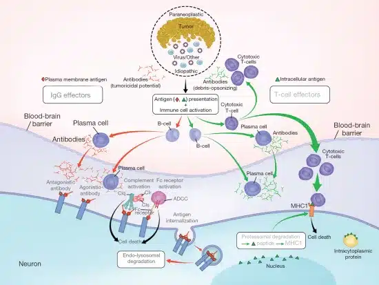
Imagine your personality suddenly changing. You become violent or sexually uninhibited. Hallucinations develop—you see things others cannot. Seizures occur. Your memory deteriorates. You become confused and disoriented. You lose consciousness. These are the devastating symptoms of autoimmune encephalitis—a rare but serious disease where the immune system mistakenly attacks brain tissue causing inflammation, neurological dysfunction, and potentially permanent brain damage. Autoimmune encephalitis is a group of disorders characterized by immune-mediated inflammation of the brain. In these diseases, autoantibodies target neuronal antigens. The antibodies bind to brain cells causing their dysfunction or destruction. Immune cells infiltrate the brain. Inflammatory mediators damage neurons. The brain inflammation—encephalitis—causes neurological symptoms. Seizures, behavioral changes, memory loss, and movement disorders develop. Without treatment, severe disability or death occurs. Autoimmune encephalitis accounts for approximately 1 to 3 percent of all encephalitis cases. The disease is more common than previously recognized. Increasing awareness has improved diagnosis. Multiple forms of autoimmune encephalitis exist. Anti-NMDA receptor encephalitis is the most common form. Other forms include anti-LGI1 encephalitis, anti-CASPR2 encephalitis, and anti-GABAb receptor encephalitis. Each form has distinctive clinical features. What makes autoimmune encephalitis particularly challenging is the variable presentation. Symptoms range from mild cognitive changes to severe psychosis and coma. Some patients recover completely with treatment. Others have permanent neurological disability. The key to good outcomes is early recognition and treatment. Modern immunotherapy has dramatically improved survival rates. In this comprehensive article, we will explore what autoimmune encephalitis is, understand how autoantibodies damage the brain, recognize neurological symptoms, learn about serious complications, understand diagnosis methods, explore available treatments, and discover management strategies for achieving neurological recovery and preventing permanent disability.
Understanding Normal Brain Function and Neurotransmission
Before we explore autoimmune encephalitis, we need to understand how neurons communicate and how antibodies disrupt this communication. The brain contains approximately 86 billion neurons. Neurons communicate through synapses—specialized connections between neurons. Neurotransmission is the process of neuronal communication. An action potential travels along the axon—the long projection of a neuron. The action potential reaches the axon terminal. Synaptic vesicles containing neurotransmitters are released. The neurotransmitters cross the synaptic cleft—the space between neurons. The neurotransmitters bind to receptors on the receiving neuron. The receptor binding causes electrical or chemical changes. These changes propagate the signal to the next neuron. Multiple neurotransmitters exist. Glutamate is the primary excitatory neurotransmitter. GABA (gamma-aminobutyric acid) is the primary inhibitory neurotransmitter. Acetylcholine is involved in movement and cognition. Dopamine regulates mood and movement. Serotonin regulates mood and emotion. Receptors are proteins on neuronal surfaces. Neurotransmitter receptors are ion channels or coupled to signaling cascades. When neurotransmitters bind, the receptors open allowing ions to flow. The ion flow changes membrane potential. The changed potential either excites or inhibits the neuron. NMDA receptors are crucial for learning and memory. These receptors bind glutamate. NMDA receptor activation is necessary for long-term potentiation—the basis of learning. LGI1 (leucine-rich glioma-inactivated 1) is involved in synapse formation. LGI1 dysfunction causes seizures and memory problems. CASPR2 (contactin-associated protein-like 2) is involved in synaptic function. CASPR2 dysfunction causes movement disorders and cognitive changes. GABA-B receptors mediate inhibitory neurotransmission. These receptors suppress neuronal firing. GABA-B receptor dysfunction causes seizures and cognitive changes. The normal balance between excitation and inhibition maintains brain function. When antibodies target these receptors, the balance is disrupted. Neuronal dysfunction results. Brain symptoms develop. Understanding receptor function helps explain why antibody-mediated receptor blockade causes specific neurological symptoms.
What is Autoimmune Encephalitis?
Autoimmune encephalitis is a group of brain inflammatory disorders where circulating autoantibodies target neuronal antigens. The antibodies bind to neurotransmitter receptors or associated proteins. The binding causes neuronal dysfunction. The antibodies also activate complement. Inflammatory mediators are generated. Microglia—brain immune cells—are activated. CD4+ and CD8+ T cells infiltrate the brain. The inflammatory environment damages neurons. Progressive neurological deterioration occurs. Multiple forms of autoimmune encephalitis are recognized based on antibody targets. Anti-NMDA receptor encephalitis is the most common form. IgG antibodies target NMDA receptors. The antibodies bind to extracellular domains of the NR1 subunit. The antibody binding prevents glutamate from acting on the receptor. NMDA receptor blockade reduces excitatory neurotransmission. Memory problems and cognitive dysfunction develop. Anti-NMDA receptor encephalitis accounts for approximately 40 to 50 percent of autoimmune encephalitis cases. Anti-LGI1 encephalitis is the second most common form. IgG antibodies target LGI1 protein. LGI1 dysfunction impairs synaptic plasticity. Memory loss and seizures are characteristic. Anti-CASPR2 encephalitis causes antibodies against CASPR2. Nystagmus (involuntary eye movements), ataxia (loss of coordination), and myoclonus (muscle jerks) are characteristic. Anti-GABAb receptor encephalitis causes antibodies against GABA-B receptors. Seizures are particularly prominent. Behavioral changes are common. Other forms of autoimmune encephalitis exist. Anti-AMPA receptor encephalitis causes cognitive and psychiatric symptoms. Anti-glycine receptor encephalitis causes rigidity and stiffness. Anti-DPPX encephalitis causes cognitive changes and movement disorders. The specific antibody determines the clinical presentation. Each antibody targets specific receptors or proteins. The dysfunction of targeted receptors causes specific neurological symptoms. Limbic encephalitis is a pattern of brain involvement. The limbic system—involved in memory and emotion—is preferentially affected. Limbic encephalitis causes memory loss, mood changes, and behavioral changes. Anti-LGI1 and anti-GABA-B antibodies commonly cause limbic encephalitis. Paraneoplastic autoimmune encephalitis is associated with cancer. Antibodies produced against cancer cells cross-react attacking brain cells. Cancers including small-cell lung cancer and ovarian cancer are associated. Finding and treating the underlying cancer helps treat autoimmune encephalitis. What causes autoimmune encephalitis is incompletely understood. Viral infections might trigger antibody production. Herpes simplex virus, Epstein-Barr virus, and other viruses have preceded disease onset. Molecular mimicry—viral antigens resembling neuronal antigens—might trigger cross-reactive antibodies. Genetic predisposition increases susceptibility. HLA types influence disease development. Female predominance in anti-NMDA receptor encephalitis suggests hormonal factors. Age influences presentation. Anti-NMDA receptor encephalitis occurs primarily in young women. Anti-LGI1 encephalitis occurs more often in older men. Anti-CASPR2 encephalitis occurs in older patients.
Recognizing Neurological Symptoms: The Changing Brain
Autoimmune encephalitis symptoms vary based on the antibody type and brain region involvement. Recognizing these symptoms prompts emergency evaluation. Psychiatric symptoms are prominent in many forms. Behavioral changes develop—personality change is striking. Patients become irritable or aggressive. Sexual disinhibition develops—inappropriate sexual behavior. Emotional instability—rapid mood swings—occurs. Depression develops. Anxiety develops. Paranoia develops—the patient believes others are conspiring against them. Paranoid ideation can be severe. Personality deterioration distresses family members. Friends report the patient is “not themselves.” Psychosis develops. Hallucinations—seeing, hearing, or feeling things others don’t—develop. Visual hallucinations are common in anti-NMDA receptor encephalitis. Auditory hallucinations occur. Tactile hallucinations develop. Delusions—false beliefs—develop. The psychotic symptoms are identical to schizophrenia. However, rapid progression distinguishes autoimmune encephalitis from schizophrenia. Cognitive changes develop. Memory impairment develops. Short-term memory deteriorates. Long-term memory can be affected. Patients cannot recall recent events. They might not recognize family members. Concentration difficulty develops. Attention span shortens. Mental processing slows. Dementia-like cognitive decline can be severe. Confusion and disorientation develop. Patients cannot recognize familiar places. They don’t know the time or date. Orientation to person—knowing their own name—can be lost. The confusion indicates severe brain dysfunction. Seizures develop in many patients. Focal seizures affecting one limb develop. Generalized seizures causing loss of consciousness develop. Status epilepticus—prolonged seizures—can occur. Seizures are medically emergent. Movement disorders develop. Dystonia—sustained muscle contractions—develops. Abnormal posturing results. Orofacial dyskinesias—involuntary facial movements—develop. Tremor develops. Myoclonus—muscle jerks—develops. Chorea—involuntary writhing movements—develops. Rigidity—stiffness—develops in some forms. Ataxia—loss of coordination—develops. Gait becomes unsteady. Walking becomes difficult. Falls occur from loss of balance. Nystagmus—involuntary eye movements—develops. Eyes move uncontrollably. Vision becomes difficult. Oculogyric crisis—eyes rolling upward—can occur. Autonomic dysfunction develops. Fever occurs despite absence of infection. Heart rate becomes abnormal—tachycardia or arrhythmias. Blood pressure becomes unstable. Sweating occurs excessively. Hypersalivation develops. Urinary retention occurs. These autonomic symptoms indicate hypothalamic involvement. Sleep disturbances develop. Insomnia develops. Sleep reversal—sleeping during day, awake at night—occurs. Nightmares are common. Consciousness levels change. Drowsiness develops. Decreased responsiveness occurs. Unresponsiveness—coma—develops in severe cases. The symptom progression is often dramatic. Patients deteriorate rapidly over days to weeks. Normal functioning becomes impossible. Hospitalization becomes necessary. Life-threatening complications develop without treatment.
Understanding Brain Inflammation: Antibody Attack and Neuronal Damage
Understanding how autoantibodies damage the brain helps explain symptom progression and treatment rationale. Autoantibody production initiates disease. For unknown reasons, B cells produce antibodies against neuronal antigens. The antibodies enter circulation. They cross the blood-brain barrier—the selective barrier protecting the brain. The antibodies reach neuronal receptors. The antibodies bind to their target receptors. NMDA receptors are blocked by anti-NMDA antibodies. The blocked receptors cannot function. Glutamate cannot activate the receptors. Excitatory neurotransmission is reduced. Memory formation requires NMDA receptor activation. NMDA receptor blockade impairs memory. LGI1 antibodies prevent LGI1 from supporting synaptic function. Synapse dysfunction occurs. Seizure threshold increases—seizures become more likely. Memory consolidation fails. CASPR2 antibodies disrupt neuronal communication. Synaptic transmission is impaired. Movement control is affected. Coordination is lost. GABA-B receptor antibodies block inhibitory neurotransmission. The blocked inhibition causes excessive neuronal firing. Seizures result. The balance between excitation and inhibition is lost. Excessive excitation predominates. Neuronal hyperactivity causes seizures and movement disorders. Complement activation follows antibody binding. C3 and C5 become activated. Inflammatory mediators are generated. The complement cascade punctures neuronal membranes. Neuronal destruction occurs. Microglia activation occurs. Microglia—the brain’s immune cells—become activated by antibodies. The activated microglia release inflammatory cytokines. TNF-alpha and interleukin-6 cause inflammation. The microglia engulf neuronal synapses. Synaptic loss occurs. T cell infiltration develops. CD8+ cytotoxic T cells infiltrate. These cells recognize antibody-coated neurons. The cytotoxic T cells kill antibody-coated neurons. Neuronal death occurs. B cell infiltration develops. B cells produce additional antibodies locally. The local antibody production amplifies inflammation. Astrocyte and oligodendrocyte activation occurs. These supporting cells become reactive. The reactive glia produce inflammatory mediators. The inflammatory environment damages neurons. Blood-brain barrier disruption occurs. Inflammatory cytokines increase BBB permeability. More immune cells enter the brain. Cerebral edema develops—the brain swells. Increased intracranial pressure develops. Herniation can occur—the brain can shift causing severe injury. The combination of antibody-mediated receptor blockade, complement activation, immune cell infiltration, and neuronal death causes severe brain damage. Progressive neurological deterioration follows. Without treatment, severe disability or death results. Understanding the mechanisms explains why immunotherapy targeting antibody production and removal is effective treatment.
Diagnosis: Recognizing Autoimmune Encephalitis
Diagnosing autoimmune encephalitis requires clinical suspicion, appropriate testing, and sometimes brain biopsy. Rapid diagnosis is crucial as treatment delays cause worse outcomes. Clinical presentation of psychiatric symptoms plus neurological symptoms in a young patient should raise suspicion. The combination is distinctive. Differential diagnoses include infectious encephalitis, schizophrenia, meningitis, and other neurological diseases. Clinical history is crucial. Doctors ask about symptom onset and progression. Rapid progression suggests autoimmune encephalitis. They ask about preceding infections. Recent upper respiratory or gastrointestinal infections precede some cases. They ask about cancer history or symptoms. Paraneoplastic autoimmune encephalitis occurs with underlying cancer. Family history of autoimmune disease is relevant. Physical examination documents neurological findings. Mental status assessment detects confusion, memory loss, or behavioral changes. Cranial nerve examination detects eye movement abnormalities. Motor examination detects movement disorders or weakness. Coordination testing detects ataxia. Gait assessment detects imbalance. Cerebrospinal fluid (CSF) analysis is crucial. Lumbar puncture obtains CSF. Elevated CSF white blood cell count indicates inflammation. Lymphocytic pleocytosis—lymphocyte predominance—suggests autoimmune or viral encephalitis. Elevated protein occurs. Normal glucose in CSF helps exclude bacterial infection. CSF oligoclonal bands—unique antibodies in CSF—support autoimmune encephalitis diagnosis. CSF autoantibody testing might detect antibodies against neuronal antigens. Some antibodies are more detectable in CSF than serum. Serum antibody testing is performed. Anti-NMDA receptor antibodies are detected by immunohistochemistry or cell-based assays. Anti-LGI1, anti-CASPR2, anti-GABA-B antibodies are similarly detected. Multiple antibody tests might be performed. Some patients are antibody-seronegative—antibodies are not detected. These patients might have non-tested antibodies or non-antibody mediated encephalitis. Brain imaging is performed. MRI brain shows characteristic findings. Temporal lobe involvement is common—limbic encephalitis. Hyperintense T2/FLAIR signals indicate inflammation. Some antibody types have characteristic imaging patterns. CT brain assesses for complications. Cerebral edema. Herniation. Increased intracranial pressure. EEG (electroencephalogram) documents brain electrical activity. Focal or generalized slowing. Seizure activity. Some antibody types show characteristic EEG patterns. “Extreme delta brush” pattern is seen in anti-NMDA encephalitis. Periodic sharp waves are seen in other forms. Brain biopsy is rarely necessary. Biopsy shows lymphocytic infiltration and neuroinflammation. Biopsy might be performed if diagnosis remains unclear. Cancer screening is important. Chest, abdomen, and pelvis imaging searches for malignancy. Gynecological examination is important in women—ovarian teratomas are associated with anti-NMDA encephalitis. Finding underlying cancer guides treatment decisions. The diagnosis of autoimmune encephalitis is confirmed when clinical features (psychiatric/neurological symptoms with rapid progression) plus CSF findings (lymphocytic pleocytosis) plus positive neuronal autoantibodies are present. Rapid diagnosis allows emergency treatment initiation.
Treatment: Immunotherapy and Brain Recovery
Autoimmune encephalitis treatment involves removing antibodies, suppressing immune response, and managing seizures. Rapid treatment is crucial for preventing permanent brain damage. Intravenous immunoglobulin (IVIG) is first-line therapy. IVIG contains pooled antibodies from thousands of donors. IVIG coats circulating pathogenic antibodies preventing their binding to neurons. IVIG modulates B cell and T cell function. IVIG is given as IV infusions over 2 to 5 days. Typical dose is 2 grams per kilogram divided over 5 days. Response occurs within days to weeks. IVIG is safer than plasmapheresis in some situations. Plasmapheresis removes circulating antibodies. Blood is filtered to remove plasma containing antibodies. Fresh frozen plasma or albumin replaces removed plasma. Antibodies are rapidly removed. Clinical improvement often follows within days. Plasmapheresis is particularly effective for antibody-mediated diseases. Five to seven daily treatments are typical. Plasmapheresis and IVIG are often used together. The combination is more effective than either alone. Rituximab depletes B cells. Rituximab is a monoclonal antibody against CD20 (B cell marker). B cell depletion reduces new antibody production. Rituximab is given IV. Clinical improvement is slower than IVIG or plasmapheresis but is sustained. Rituximab prevents relapse. High-dose corticosteroids suppress immune response. Methylprednisolone 1 gram IV daily for 3 to 5 days. Oral prednisone follows. Corticosteroids reduce immune cell activation. The immunosuppression reduces inflammation. Corticosteroids are often used with other therapies. Seizure control is crucial. Antiepileptic drugs prevent seizures. Levetiracetam, valproate, or other drugs are used. Multiple antiepileptic drugs might be necessary. Lorazepam or other benzodiazepines treat acute seizures. Status epilepticus is a medical emergency. ICU-level care is often necessary. Mechanical ventilation supports breathing if consciousness is impaired. Sedation controls agitation. Intracranial pressure monitoring prevents herniation. Cooling blankets manage fever. Plasmapheresis combined with corticosteroids and rituximab is standard treatment. This triple therapy produces remission in most patients. Approximately 80 percent of anti-NMDA encephalitis patients respond to treatment. Response rates vary for other antibody types. Early treatment predicts better outcomes. Treatment delays allow more neuronal damage increasing permanent disability risk. Some patients have severe encephalitis resistant to initial treatment. Cyclophosphamide is added for refractory disease. Cyclophosphamide produces deeper immune suppression. Additional immune modulatory therapies are sometimes used. The key is rapid initiation of multiple immune therapies. Combination treatment produces best outcomes.
Living with Autoimmune Encephalitis: Recovery and Long-Term Management
Living with autoimmune encephalitis requires recovery support, seizure management, and neurological rehabilitation. Many patients recover substantially with aggressive treatment. However, recovery is often prolonged. Cognitive recovery develops over weeks to months. Memory usually returns gradually. Personality normalization occurs. Behavioral changes resolve. Some residual cognitive changes persist. Concentration might not fully recover. Memory gaps might remain. However, most patients achieve functional recovery. Physical recovery develops. Movement disorders resolve. Coordination improves. Gait normalizes. Muscle strength returns. Most patients eventually achieve normal physical function. Psychological recovery is important. Patients often struggle with memory of psychiatric symptoms. Psychosis and behavioral changes are distressing to recall. Counseling helps process trauma. Depression sometimes develops. Antidepressants help some patients. Support groups provide understanding from others. Seizure management continues. Some patients become seizure-free. Others require lifelong antiepileptic drugs. Multiple antiepileptic drug combinations might be necessary. Regular monitoring ensures seizure control. Continued immunosuppression prevents relapse. Some patients require extended corticosteroid therapy. Long-term rituximab or other immunosuppression might be necessary. Gradually tapering immunosuppression occurs as remission is stable. Complete tapering of immunosuppression can occur. However, relapse can occur requiring reinitiation of therapy. Regular neurological assessment monitors recovery. Neuropsychological testing assesses cognitive recovery. Imaging reassesses brain. EEG confirms seizure control. Rehabilitation helps maximize recovery. Cognitive rehabilitation helps memory and concentration recovery. Physical therapy helps movement recovery. Occupational therapy helps daily functioning. Speech therapy helps if language is affected. Neuropsychological counseling addresses emotional effects. Work and school return requires planning. Gradual return is usually necessary. Cognitive changes might affect work performance. Some patients require work accommodations. Others require disability. Education about autoimmune encephalitis helps understanding. Patients should understand the disease. They should understand treatment. They should understand recovery expectations. Some residual effects might persist. Most achieve substantial functional recovery. Mental health support continues. Regular follow-up ensures sustained remission. Annual neurological assessment continues indefinitely. Most patients achieve normal or near-normal life. However, some have permanent neurological effects. Memory problems might persist. Cognitive changes might persist. Personality changes might persist. Seizures might recur. However, with appropriate treatment and rehabilitation, quality of life is usually good.
Frequently Asked Questions (FAQs)
Q1: Can autoimmune encephalitis be cured?
Autoimmune encephalitis cannot be cured in the sense that the underlying autoimmune dysregulation is permanent. However, immunotherapy effectively suppresses disease. Most patients achieve remission with immunosuppressive treatment. The antibodies decrease. Inflammation resolves. Neurological symptoms improve. Most patients recover substantially. Some require continued immunosuppression to maintain remission. Relapse can occur if treatment is discontinued prematurely. With appropriate long-term management, most patients maintain remission and normal neurological function.
Q2: Is autoimmune encephalitis fatal?
Autoimmune encephalitis can be fatal if untreated or if treatment is delayed. Severe cases develop status epilepticus—prolonged seizures. Cerebral edema and increased intracranial pressure develop. Herniation can occur. Respiratory failure can develop. Without treatment, mortality rate approaches 40 to 50 percent. However, with prompt immunotherapy, mortality rate has decreased to less than 10 percent. Early diagnosis and aggressive treatment prevent most deaths.
Q3: Why do antibodies attack the brain in autoimmune encephalitis?
The reason why the immune system produces antibodies against neuronal antigens is incompletely understood. Viral infections might trigger antibody production through molecular mimicry. Neuronal antigens might resemble viral antigens. Immune response against viruses cross-reacts attacking brain cells. Genetic predisposition increases susceptibility. Some people genetically predisposed to autoimmunity develop autoimmune encephalitis when exposed to environmental triggers. Cancer-associated autoimmune encephalitis results from antibodies produced against cancer cells that cross-react against brain.
Q4: How long does recovery from autoimmune encephalitis take?
Recovery time varies dramatically. Some patients show rapid improvement within days of starting immunotherapy. Others improve gradually over weeks to months. Most substantial improvement occurs in the first 3 to 6 months. However, full neurological recovery can take a year or longer. Some residual neurological effects persist. Memory problems might partially persist. Cognitive changes might partially persist. However, most patients achieve functional recovery allowing return to work or school within 6 to 12 months.
Q5: Can autoimmune encephalitis recur?
Yes, relapse can occur in approximately 10 to 20 percent of patients. Relapse usually occurs within the first year after initial treatment. Some patients have multiple relapses. Others relapse only once. Maintaining appropriate immunosuppression reduces relapse risk. Some experts recommend prolonged immunosuppression in all patients. Others gradually taper therapy in remission-remission. Close monitoring continues indefinitely. Regular neurology follow-up detects early relapse signs. Prompt retreatment of relapse prevents severe neurological damage.
Key Takeaways
Autoimmune encephalitis is a group of brain inflammatory disorders characterized by autoantibodies targeting neuronal antigens. Anti-NMDA receptor encephalitis is the most common form. Psychiatric symptoms including psychosis and behavioral changes are prominent. Neurological symptoms including seizures, memory loss, and movement disorders develop. Rapid disease progression distinguishes autoimmune from infectious encephalitis. CSF lymphocytic pleocytosis and positive neuronal autoantibodies confirm diagnosis. IVIG, plasmapheresis, and rituximab are first-line immunotherapy. Corticosteroids and seizure management support treatment. Rapid diagnosis and early treatment are crucial for good outcomes. Approximately 80 to 90 percent response rate with modern immunotherapy. Mortality rate decreased to less than 10 percent with prompt treatment. Recovery is often gradual over weeks to months. Most patients achieve substantial functional recovery. Some residual neurological effects might persist. Relapse occurs in 10 to 20 percent of patients. Long-term neurological follow-up continues indefinitely. With appropriate treatment and management, most patients achieve good quality of life.
References
- World Health Organization (WHO). “Autoimmune Encephalitis and Neurological Autoimmunity.” Retrieved from https://www.who.int/
- American Academy of Neurology. “Autoimmune Encephalitis: Clinical Guidelines.” Retrieved from https://www.aan.com/
- Mayo Clinic. “Autoimmune Encephalitis: Causes and Treatment.” Retrieved from https://www.mayoclinic.org/
- Cleveland Clinic. “Autoimmune Encephalitis: Complete Information.” Retrieved from https://my.clevelandclinic.org/
- National Institute of Neurological Disorders and Stroke. “Encephalitis.” Retrieved from https://www.ninds.nih.gov/
- American Brain Foundation. “Autoimmune Encephalitis Patient Resources.” Retrieved from https://www.americanbrainfoundation.org/
Related Articles on ObserverVoice.com
Explore more health and science topics on our platform:
- Understanding Autoimmune Diseases: When Your Immune System Attacks Itself
- The Brain: Function, Structure, and Neurological Health
- Seizures and Epilepsy: Understanding Neurological Convulsions
- Psychiatric Disorders: Understanding Mental Health Conditions
- Memory and Cognition: Understanding Brain Function
- Living with Chronic Illness: Adaptation and Support Strategies
Disclaimer
This article adapts publicly available information from WHO sources. This content is for informational and educational purposes only and does not constitute medical advice. [ObserverVoice.com] is a news and information platform — not a healthcare provider. If you suspect you have autoimmune encephalitis, experiencing psychiatric symptoms, seizures, or neurological changes, seek emergency medical care immediately. Autoimmune encephalitis can progress rapidly causing severe brain damage. Early diagnosis and prompt immunotherapy are crucial for preventing permanent disability. Always seek immediate medical attention for emergency neurological symptoms. Licensed healthcare specialists must diagnose and treat autoimmune encephalitis.
Observer Voice is the one stop site for National, International news, Sports, Editor’s Choice, Art/culture contents, Quotes and much more. We also cover historical contents. Historical contents includes World History, Indian History, and what happened today. The website also covers Entertainment across the India and World.

