

Autoimmune Encephalitis: When the Brain Becomes the Target of an Immune Attack

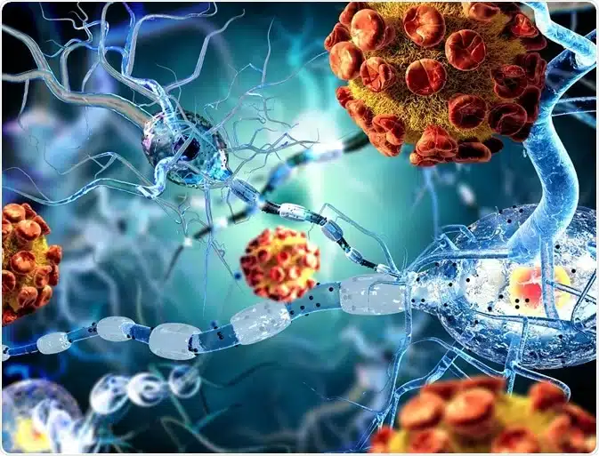
Autoimmune Encephalitis is a rare but serious disease in which the immune system abnormally produces antibodies against proteins and receptors in the brain. These antibodies attack brain cells and neurons. Inflammation of brain tissue occurs. Encephalitis is inflammation of the brain. Progressive neurological dysfunction results. Seizures develop. Psychiatric symptoms develop. Cognitive decline occurs. Memory loss occurs. Behavioral changes occur. Autoimmune Encephalitis encompasses multiple distinct forms. Each with different antibody targets. NMDA receptor encephalitis. Most common form. Anti-NMDA receptor antibodies. AMPA receptor encephalitis. Anti-AMPA receptor antibodies. GABA receptor encephalitis. Anti-GABA receptor antibodies. LGI1 encephalitis. Anti-leucine-rich glioma-inactivated 1 antibodies. CASPR2 encephalitis. Anti-contactin-associated protein 2 antibodies. Other forms. Anti-dopamine-2 receptor antibodies. Anti-IgLON5 antibodies. Others. The antibodies disrupt normal brain function. NMDA receptors mediate synaptic transmission. Blocking NMDA receptors impairs cognition. Impairs memory. Causes psychiatric symptoms. AMPA receptors essential for synaptic plasticity. Blocking causes cognitive dysfunction. GABA receptors inhibitory. Blocking causes seizures. Loss of inhibition. Hyperexcitability. The brain inflammation causes multiple symptoms. Seizures from neuronal hyperexcitability. Psychiatric manifestations from limbic involvement. Cognitive decline from cortical involvement. Movement disorders from basal ganglia involvement. Autonomic dysfunction from brainstem involvement. Autoimmune Encephalitis affects approximately one to three per one million population annually. The disease is rare. However, increasingly recognized. Improved diagnostic methods. Better awareness. Previously unrecognized cases now identified. The disease affects all age groups. Most common in young adults. Peak age twenty to thirty years. However, can develop in children. Can develop in elderly. Female predominance. Women more frequently affected. Approximately seventy percent of cases. Autoimmune Encephalitis is caused by abnormal immune activation producing autoimmune antibodies. Exact triggers unknown. Paraneoplastic. Associated with malignancy in some. Malignancy triggers antibodies. Non-paraneoplastic. No malignancy. Triggered by other factors. Genetic predisposition. Environmental trigger possibly. Infections. Vaccinations. HLA associations. Early diagnosis and immediate immunosuppression are crucial for preventing permanent neurological damage. With early aggressive treatment, remission is achievable. Neurological recovery possible. Permanent disability prevented. Understanding Autoimmune Encephalitis helps with early recognition and appropriate urgent management to prevent severe brain damage and permanent disability.
How Do Autoimmune Antibodies Cause Brain Inflammation and Neurological Dysfunction?
To understand Autoimmune Encephalitis, we need to learn about the brain and synaptic transmission. The brain is the control center. Billions of neurons. Neurons communicate through synapses. Neurotransmitters transmitted across synapses. Receptors on postsynaptic cells receive signals. Synaptic transmission essential for normal brain function. Cognition. Memory. Emotion. Movement. Sensation. Neurotransmitter receptors include NMDA receptors. AMPA receptors. GABA receptors. Glutamate receptors. Excitatory. Mediate excitatory transmission. GABA receptors. Inhibitory. Mediate inhibitory transmission. Balance of excitation and inhibition essential. Appropriate brain function. In Autoimmune Encephalitis, autoimmune antibodies are produced. Antibodies against specific brain antigens. NMDA receptors. AMPA receptors. GABA receptors. LGI1. CASPR2. Others. The antibodies circulate in blood. Cross blood-brain barrier. Accessible to brain tissue. Antibodies bind to receptors. Block receptor function. Or cross-link receptors. Internalization of receptors. Receptors removed from surface. Degraded intracellularly. Loss of receptor density. Receptor dysfunction. Synaptic transmission impaired. The antibodies activate complement. Classical complement pathway. C3 deposition. MAC formation. Membrane attack complex. Neuronal cell membranes damaged. Neuron death. Cell lysis. The antibodies also activate cellular immunity. T cell recruitment. T cells infiltrate brain. Produce inflammatory cytokines. Interleukin-2, interleukin-6, TNF-alpha. Interferon-gamma. These inflammatory mediators amplify inflammation. More immune cell infiltration. More cytokine production. Inflammatory cascade. Microglial activation. Microglia are brain macrophages. Activation produces pro-inflammatory cytokines. Inflammatory mediators. Neuroinflammation amplifies. Astrocyte activation. Astrocytes produce cytokines. Chemokines. Reactive oxygen species. Free radicals. Oxidative stress. Neuronal damage. Blood-brain barrier disruption. Barrier function impaired. More immune cells infiltrate. Edema. Brain swelling. Increased intracranial pressure. Herniation risk. The brain inflammation is diffuse. Limbic encephalitis. Medial temporal lobe predominantly. Amygdala. Hippocampus. Involved. Memory impairment. Behavioral changes. Seizures. Cortical involvement. Frontal and temporal cortex. Cognitive dysfunction. Psychiatric symptoms. Behavioral abnormalities. Basal ganglia involvement. Movement disorders. Dystonia. Dyskinesia. Parkinsonism. Brainstem involvement. Autonomic dysfunction. Autonomic instability. Autonomic crisis. Temperature dysregulation. Autonomic storm. Life-threatening. The neuronal dysfunction results from receptor loss and synaptic transmission impairment. NMDA receptor dysfunction. Hypofunction. Schizophrenia-like symptoms. Cognitive decline. Memory impairment. Psychosis. AMPA receptor dysfunction. Loss of synaptic plasticity. Memory deficits. Cognitive dysfunction. GABA receptor dysfunction. Loss of inhibition. Hyperexcitability. Seizures. Status epilepticus. LGI1 dysfunction. Seizures. Memory dysfunction. Faciobrachial dystonic seizures. Characteristic. CASPR2 dysfunction. Neuromyotonia. Muscle stiffness. Involuntary contractions. Seizures. The progressive nature of the disease. Initial symptoms mild. Progressive worsening. Over days to weeks. Severe dysfunction. Status epilepticus. Autonomic crisis. Coma. Death without treatment. Understanding the antibody-mediated mechanisms has led to development of immunotherapy targeting B cells and plasma cells.
What Are the Main Symptoms and Signs of Autoimmune Encephalitis?
Autoimmune Encephalitis causes diverse neurological and psychiatric symptoms depending on the antibody type and brain areas involved. Psychiatric symptoms often present first. Personality change. Behavioral abnormality. Unusual behavior. Out of character. Profound personality shift. Psychiatric manifestations. Psychosis. Hallucinations. Auditory hallucinations. Hearing voices. Visual hallucinations. Seeing things. Delusions. False beliefs. Paranoia. Persecutory delusions. Mania. Elevated mood. Excessive energy. Decreased need for sleep. Risky behavior. Grandiosity. Depression. Depressed mood. Suicidal ideation. Suicide attempt. Catatonia. Muteness. Waxy flexibility. Posturing. Complete unresponsiveness. Bizarre behavior. Agitation. Aggressive. Violence. Aggressiveness. Unprovoked. Unusual for person. Anxiety. Panic attacks. Obsessive compulsive symptoms. Compulsions. Intrusive thoughts. Cognitive symptoms develop. Memory loss. Amnesia. Inability to form new memories. Anterograde amnesia. Inability to recall recent events. Difficulty concentrating. Attention difficulty. Slowed thinking. Mental slowness. Difficulty with complex tasks. Word-finding difficulty. Aphasia. Language dysfunction. Seizures develop. Generalized tonic-clonic seizures. GTCS. Full-body seizure. Loss of consciousness. Tonic phase. Clonic phase. Status epilepticus. Prolonged seizures. Medical emergency. Focal seizures. Partial seizures. Specific brain region. May progress to generalized. Faciobrachial dystonic seizures. Characteristic of LGI1. Mouth grimacing. Arm dystonia. Brief. Stereotyped. Repeated. Seizure frequency. Intractable seizures. Frequent. Daily. Multiple daily. Difficult to control. Movement disorders. Dystonia. Abnormal posture. Involuntary muscle contraction. Twisted postures. Painful. Dyskinesia. Involuntary abnormal movements. Chorea. Athetosis. Ballism. Parkinsonism. Stiffness. Bradykinesia. Tremor. Rigidity. Hypokinesia. Reduced movement. Reduced speech volume. Reduced facial expression. Muscle rigidity. Stiffness of muscles. Neuromyotonia. Muscle stiffness. Involuntary muscle contractions. Fasciculations. Visible muscle twitching. Ataxia. Loss of coordination. Unsteady gait. Incoordination of movements. Falls. Autonomic symptoms. Fever. Elevated temperature. May persist. Autonomic dysregulation. Dysautonomia. Tachycardia. Rapid heart rate. Blood pressure instability. Hypertension alternating with hypotension. Flushing. Sweating. Profuse sweating. Temperature dysregulation. Hypothermia. Hyperthermia. Difficulty maintaining temperature. Respiratory dysfunction. Tachypnea. Rapid breathing. Hypoventilation. Shallow breathing. Apnea. Breathing cessation. Requiring mechanical ventilation. Sleep disturbance. Insomnia. Inability to sleep. Hypersomnia. Excessive sleeping. REM sleep behavior disorder. Acting out dreams. Autonomic storm. Life-threatening. Extreme fever. Extreme tachycardia. Extreme hypertension. Profound sweating. Autonomic instability. Medical emergency. Headache. Persistent headache. Meningismus. Neck stiffness. Photophobia. Light sensitivity. Phonophobia. Sound sensitivity. Altered consciousness. Confusion. Disorientation. Difficulty with time orientation. Place orientation. Person orientation. Stupor. Lethargy. Extreme sleepiness. Difficult to arouse. Coma. Unresponsiveness. Inability to arouse. Mutism. Inability to speak. Selective mutism. Speaks minimally. Swallowing difficulty. Dysphagia. Aspiration risk. Incontinence. Loss of bladder control. Loss of bowel control. The progression is typically rapid. Days to weeks. Some slower. Weeks to months. Without treatment, progressive worsening. Severe disability. Coma. Death. Early recognition crucial.
How is Autoimmune Encephalitis Detected and Diagnosed?
Autoimmune Encephalitis is diagnosed through clinical presentation, laboratory testing for antibodies, and imaging studies showing brain inflammation. Early diagnosis is crucial. Clinical history is important. Acute neuropsychiatric symptoms. Seizures. Behavioral changes. Personality changes. Memory loss. Rapid progression. Progressive worsening. Often preceded by viral illness. URI. Fever. Systemic illness. Or psychiatric change first. Seizures later. Physical examination findings. Altered mental status. Confusion. Disorientation. Abnormal behavior. Seizures during examination. Abnormal movements. Dystonia. Dyskinesia. Ataxia. Signs of meningitis. Neck stiffness. Kernig sign. Brudzinski sign. Fever. Abnormal vital signs. Tachycardia. Hypertension. Tachypnea. Cerebrospinal fluid analysis. CSF lumbar puncture. Essential for diagnosis. Pleocytosis. Elevated white blood cells. Usually lymphocytic. Lymphocytes predominant. Not PMN predominant. Protein elevation. CSF protein elevated. Glucose. CSF glucose usually normal. Elevated protein with normal glucose. Characteristic. Not bacterial meningitis. CSF cultures. Negative. Rules out bacterial infection. Viral cultures. Usually negative. Gram stain. Negative. Rules out bacterial. PCR for common viruses. Usually negative. Rules out viral encephalitis. Serum antibody testing. Anti-NMDA receptor antibodies. ELISA test. IgG and IgM. IgG more specific. Anti-AMPA receptor antibodies. Anti-GABA receptor antibodies. Anti-LGI1 antibodies. Anti-CASPR2 antibodies. Others. Highly specific. Presence confirms diagnosis. CSF antibody testing. More specific than serum. CSF antibodies indicate intrathecal synthesis. Brain-specific antibodies. More specific. Serum and CSF negative. Still consider autoimmune. Clinical diagnosis. Trial of immunotherapy. Response supports diagnosis. Brain MRI. Shows brain inflammation. FLAIR sequences. T2 sequences. Signal abnormalities. Typically T2/FLAIR hyperintensities. Medial temporal lobes. Limbic encephalitis. Focal or multifocal. Enhancement with gadolinium. Suggests inflammation. Blood-brain barrier breakdown. Gray matter predominant. White matter usually spared. MRI may be normal early. Repeat MRI. Evolution of findings. PET scan. FDG-PET. Shows metabolic changes. Hypermetabolism in inflamed areas. Useful if MRI normal. CT head. Rules out complications. Hemorrhage. Edema. Herniation. Baseline. Not specific. EEG findings. Electroencephalography. Shows abnormalities. Generalized slowing. Background slowing. Focal slowing. Seizure activity. Epileptiform discharges. Spikes. Sharp waves. Status epilepticus pattern. Extreme delta brush. EDD. Extreme delta brush. Bursts of delta with superimposed faster activity. Characteristic of anti-NMDA. Very specific finding. Long-duration monitoring. Video EEG. Captures seizures. Localizes ictal onset. Paraneoplastic workup. If suspected. Cancer screening. CT chest. CT abdomen. Ultrasound. PET scan. Colonoscopy. Gynecology exam. Age-appropriate cancer screening. The combination of clinical presentation with psychiatric and neurological symptoms, seizures, CSF pleocytosis, brain MRI showing inflammation, positive antibody testing, and exclusion of infection confirms Autoimmune Encephalitis diagnosis. Early diagnosis allows immediate immunotherapy.
What Health Complications Do People with Autoimmune Encephalitis Face?
People with Autoimmune Encephalitis face serious complications from brain inflammation and neuronal dysfunction. The complications depend on disease severity and timing of treatment. Status epilepticus is the most immediately life-threatening complication. Prolonged seizures. Continuous. Lasting thirty minutes or more. Refractory status epilepticus. Fails initial therapy. Life-threatening. Breathing difficulties. Respiratory failure. Aspiration risk. Metabolic acidosis. Lactic acidosis. Hyperthermia. Extreme fever. Brain damage. Neuronal death. Permanent brain injury. Cerebral edema. Brain swelling. Increased intracranial pressure. Herniation. Brainstem herniation. Transtentorial herniation. Death. Without urgent treatment. Mortality high. Autonomic crisis. Severe dysautonomia. Extreme fever. One hundred four degrees Fahrenheit or higher. Extreme tachycardia. Heart rate two hundred or higher. Hypertensive crisis. Blood pressure two hundred or higher. Profuse sweating. Cardiovascular collapse. Shock. Renal failure. Acute kidney injury. Rhabdomyolysis. Muscle breakdown. Myoglobin release. Kidney damage. Disseminated intravascular coagulation. DIC. Thrombosis. Bleeding. Death. Medical emergency. Coma. Prolonged unconsciousness. Minimal responsiveness. Unresponsiveness to stimuli. Vegetative state. Minimal conscious state. Coma may be reversible. However, if prolonged. Brain damage. Permanent consciousness. Death. Mechanical ventilation dependence. Prolonged intubation. Ventilator dependence. Difficulty weaning. Chronic respiratory support. Aspiration. Food or secretions into lungs. Aspiration pneumonia. Infection. Serious infection. Mechanical ventilation complications. Ventilator-associated pneumonia. VAP. Sinusitis. Otitis media. Otitis externa. Infection risk. Immunosuppression. From immunotherapy. Infection susceptibility. Opportunistic infections. PCP. CMV. Fungal infections. Bacterial infections. Sepsis. Septic shock. Death. Seizure-related injuries. Head trauma from falls. Fractures. Concussion. Subdural hematoma. Epidural hematoma. Serious intracranial injury. SIADH. Syndrome of inappropriate antidiuretic hormone. Hyponatremia. Low sodium. Seizures from electrolyte abnormality. Encephalopathy. Brain edema. Death. Cardiovascular complications. Myocarditis. Heart muscle inflammation. Heart failure. Arrhythmias. Sudden cardiac death. Cardiomyopathy. Reduced ejection fraction. Pulmonary edema. Respiratory failure. Rhabdomyolysis complications. Acute kidney injury. Dialysis need. Permanent renal failure. Death. Metabolic complications. Hyperthermia complications. Heat stroke. Organ damage. Disseminated intravascular coagulation. Death. Psychosis complications. Suicidal behavior. Suicide attempt. Successful suicide. Mortality from suicide. Self-harm. Intentional injury. Serious. Violent behavior. Injury to self. Injury to others. Legal consequences. Permanent cognitive impairment. Memory loss. Persistent. Dementia-like. Difficulty with new learning. Difficulty with complex tasks. Reduced work capacity. Disability. Permanent neurological deficits. Residual seizures. Ongoing seizures. Despite treatment. Intractable epilepsy. Movement disorder. Dystonia. Dyskinesia. Persistent. Parkinsonism. Chronic. Cerebellar dysfunction. Ataxia. Permanent. Incoordination. Falls. Disability. Mood disorder. Depression. Anxiety. PTSD. Post-traumatic stress. From ICU experience. From severe illness. Personality change. Behavioral abnormality. Persistent. Different from before. Relationship strain. Family impact. Cognitive dysfunction. Permanent. Persistent. Work difficulty. School difficulty. Disability. Without early aggressive immunotherapy, permanent brain damage likely. With early treatment, recovery possible. However, some persistent effects possible. Most recover well with early treatment.
What Treatments Help People with Autoimmune Encephalitis?
Treatment for Autoimmune Encephalitis focuses on urgent immunosuppression to halt immune attack and reduce brain inflammation. Immediate treatment is critical. First-line immunotherapy. Corticosteroids. High-dose methylprednisolone. IV corticosteroids. Pulse therapy. One gram daily. Three to five days. Rapid anti-inflammatory effect. Suppresses immune activation. Then transition to oral prednisone. One to two milligrams per kilogram. Taper over weeks to months. Long-term low-dose maintenance. Intravenous immunoglobulin. IVIG. Polyvalent immunoglobulin. From pooled human plasma. Modulates immune response. Blocks pathogenic antibodies. Decreases cytokine production. Multiple mechanisms. High-dose IVIG. Two grams per kilogram. Over three to five days. Divided doses. Repeat monthly if needed. Shows benefit. Often combined with corticosteroids. Plasmapheresis. Plasma exchange. Removes circulating antibodies. Removes pathogenic immunoglobulins. Rapid effect. Five to seven exchanges. Over one to two weeks. Daily exchanges. Alternates with rest days. Removes antibodies directly. Effective. Mechanical removal. Combined with immunosuppression. Second-line immunotherapy. Rituximab. B cell depletion. Monoclonal antibody. CD20 targeting. B cell destruction. Reduces antibody production. Used for refractory cases. Used if first-line inadequate. Two infusions. One week apart. Four hundred milligrams per dose. Or weight-based dosing. Shows benefit. Used increasingly. Mycophenolate mofetil. MMF. Immunosuppressive agent. T cell and B cell suppression. Reduces antibody production. Used with corticosteroids. Azathioprine. Immunosuppressive agent. Long-term use. Used as maintenance. After acute phase. Cyclophosphamide. Cytotoxic agent. Used for severe refractory cases. B cell suppression. Potent immunosuppression. Serious side effects. Cancer risk. Bladder toxicity. Reserved for severe. Long-term management. Tacrolimus. Calcineurin inhibitor. T cell suppression. Used as maintenance. Data limited. Some benefit reported. Seizure management. Anticonvulsants essential. Levetiracetam. First-line. Valproic acid. Phenytoin. Lacosamide. Clobazam. Benzodiazepines. Acute seizure management. Lorazepam. Diazepam. Status epilepticus. Rescue medication. Fosphenytoin. IV antiepileptic. Phenobarbital. Second-line antiepileptic. Multiple agents. Polypharmacy common. Goal seizure control. Refractory seizures. Multiple agents. High doses. Vagal nerve stimulator. For refractory. Supportive care. Mechanical ventilation if respiratory failure. Intubation. Endotracheal tube. ICU monitoring. Continuous monitoring. Autonomic support. Cooling measures if fever. Cooling blankets. Cold saline. Antipyretics. Acetaminophen. NSAIDs if not contraindicated. Aggressive fever management. Autonomic medications. For dysautonomia. Beta-blockers. Propranolol. Atenolol. For tachycardia. Antihypertensives. Vasodilators. For hypertension. Labetalol. Nicardipine. Fluid management. IV fluids. Support. Electrolyte management. Sodium supplementation. If hyponatremia. Careful monitoring. SIADH treatment. Fluid restriction. Hypertonic saline. If severe hyponatremia. Neuroimaging. Repeat MRI. Document response. Monitor inflammation. CT if complications. Hemorrhage. Edema. Herniation. Cancer screening. If paraneoplastic suspected. Age-appropriate screening. Imaging. Blood tests. Colonoscopy. With appropriate urgent immunotherapy, most achieve remission or significant improvement. Recovery often dramatic. Seizures stop. Psychiatric symptoms resolve. Cognition improves. Early treatment critical. Best outcomes.
Living with Autoimmune Encephalitis
Living with Autoimmune Encephalitis requires ongoing immunosuppression, seizure management, cognitive rehabilitation, and psychological adjustment to a disease with potential for permanent neurological effects. For people surviving acute Autoimmune Encephalitis, recovery is often remarkable. Dramatic improvements. Return to baseline. However, some residual effects possible. Ongoing management important. Long-term immunosuppression. Corticosteroid continuation. Low-dose maintenance. Taper gradually. But often lifelong low-dose needed. Discontinuation risks relapse. Immunosuppressive agents. Mycophenolate mofetil. Long-term continuation. Azathioprine. Maintenance therapy. Regular monitoring essential. Antibody titers. Monitor if available. Declining titers. Good sign. Persistent antibodies. Relapse risk. Rising titers. Relapse warning. Repeat immunotherapy. Brain MRI. Periodic imaging. Document resolution. Monitor for recurrence. EEG. Periodic EEG. Assess seizure control. Background activity. Resolution of abnormalities. Seizure management. Anticonvulsants. Long-term continuation. Seizure control maintenance. Multiple agents. Often needed. Seizure threshold. Seizure precautions. Avoid triggers. Sleep deprivation. Stress. Flashing lights. Physical activity. Regular moderate exercise. Low-impact. Walking. Swimming. Cycling. Avoid heavy exertion. Strenuous exercise. Overheating risk. Infection prevention. Immunosuppression increases infection risk. Vaccination. Annual flu vaccine. Pneumococcal vaccine. Meningococcal vaccine. Other vaccines. Prophylactic antibiotics. Sometimes used. TMP-SMX for PCP prophylaxis. Hand hygiene. Avoid infections. Stress management. Stress may trigger relapse. Meditation and relaxation. Yoga. Regular exercise. Counseling. Reduces stress. Sleep quality. Important for recovery. Regular sleep schedule. Adequate sleep. Sleep hygiene. Avoid sleep deprivation. Medication side effects. Corticosteroid side effects. Hypertension. Hyperglycemia. Osteoporosis. Infection risk. Immunosuppressive side effects. GI symptoms. Liver toxicity. Leukopenia. Anticonvulsant side effects. Ataxia. Somnolence. Cognitive impairment. Some resolve with time. Require adjustment. Nutrition management. Balanced diet. Adequate calories. Adequate protein. Calcium and vitamin D. Bone health. Corticosteroid-induced osteoporosis. Vitamin supplementation. Vitamins lost with disease. Rehabilitation. Cognitive rehabilitation. Memory exercises. Concentration exercises. Speech therapy. If aphasia. Physical therapy. If movement disorders. Ataxia. Dystonia. Dyskinesia. Occupational therapy. Activities of daily living. Adapting to disabilities. Psychological support. PTSD. From ICU experience. From severe illness. Trauma counseling. PTSD therapy. Depression. From disease. From disability. From residual effects. Antidepressants. Psychotherapy. Anxiety. Anxiety treatment. Antianxiety medications. Counseling. Support groups. Others with autoimmune encephalitis. Share experiences. Coping strategies. Online communities. Work and school adjustments. Cognitive dysfunction. May affect work capacity. School performance. Some need accommodations. Reduced hours. Modified duties. School IEPs. Seizures. Unpredictable. Restrictions. Driving restrictions. Until seizure-free. State-dependent. Requirements vary. Dating and relationships. Explaining condition. Disease rarity. Unusual presentation. Understanding from partners. Seizures. Medication side effects. Cognitive effects. Sexual dysfunction. From disease. From medications. Communication helps. Support. Pregnancy. Women with autoimmune encephalitis can become pregnant. Requires careful planning. Antiepileptic drugs. Some teratogenic. Medication adjustment. Risk of relapse in pregnancy. Careful monitoring. Fetal monitoring. Usually successful with precautions. Regular medical follow-up. Neurology follow-up. Regular assessment. Seizure control. Medication side effects. Relapse assessment. Immunology follow-up. Immunosuppression monitoring. Infection risk. Psychiatric follow-up. If residual psychiatric symptoms. Support. Family education. Understanding disease. Understanding recovery. Understanding relapse risk. Explaining seizures. Seizure first aid. CPR. Seizure precautions. Support with management. Emotional support. With appropriate urgent immunotherapy, ongoing seizure management, cognitive and physical rehabilitation, psychological support, stress management, infection prevention, medication compliance, family and social support, and regular medical monitoring, most Autoimmune Encephalitis survivors achieve good recovery and maintain reasonable quality of life despite the serious nature of this autoimmune brain disease.
Frequently Asked Questions About Autoimmune Encephalitis
FAQ 1: Can Autoimmune Encephalitis be cured? Autoimmune Encephalitis cannot be cured. The underlying autoimmune process is chronic. However, remission is achievable. Complete remission with no symptoms. Antibodies undetectable. Some relapse. Reactivation of disease. Usually responds to renewed immunotherapy. Long-term immunosuppression often necessary. Prevention of relapses important. Most controlled well with ongoing therapy. Good long-term outcomes possible.
FAQ 2: What is the prognosis for Autoimmune Encephalitis? Prognosis depends on timing of diagnosis and treatment. Early diagnosis. Immediate immunotherapy. Best outcomes. Most recover well. Return to baseline. Some residual effects. Later diagnosis. Delays in treatment. More permanent damage. Worse prognosis. With modern immunotherapy. Mortality decreased. Most survive. Morbidity remains concern. Permanent disability possible. However, most recover functional. Good prognosis with early treatment.
FAQ 3: Will Autoimmune Encephalitis recur? Autoimmune Encephalitis can recur. Relapse rate variable. Approximately twenty to thirty percent. Usually within first two years. However, can occur later. Relapse indicated by returning symptoms. Psychiatric symptoms. Seizures. Cognitive decline. Antibody titers rising. Usually responds to renewed immunotherapy. Early recognition important. Regular monitoring. Long-term prophylactic immunosuppression. Reduces relapse risk. Important.
FAQ 4: Can the psychiatric symptoms from Autoimmune Encephalitis persist? Psychiatric symptoms usually resolve with immunotherapy. However, some may persist. Residual mood changes. Personality change. Cognitive dysfunction. Persistent memory problems. Persistent concentration difficulty. Cognitive rehabilitation. Helps. Psychotherapy. Helps with adjustment. Antidepressants. For persistent depression. Most psychiatric symptoms resolve. However, support important.
FAQ 5: Are there new treatments being developed for Autoimmune Encephalitis? Yes, research ongoing. Better antibody testing. More specific. Faster diagnosis. New immunosuppressants. More targeted. Fewer side effects. B cell and plasma cell targeted therapy. More specific. CAR-T cell therapy. Targeting T cells. Experimental. Tolerance induction. Restoring immune tolerance. Preventing relapse. Biomarker research. Predicting treatment response. Predicting prognosis. Gene therapy. Correcting immune defect. Experimental. Better treatments anticipated. Improved outcomes expected.
References and Further Reading
For more information about Autoimmune Encephalitis, you can visit several trusted and authoritative sources providing detailed information for patients and families dealing with this rare autoimmune brain disease. The World Health Organization at WHO.int provides comprehensive information about Autoimmune Encephalitis and related neurological diseases. The Autoimmune Encephalitis Alliance at AutoimmuneEncephalitis.org provides patient education and support specifically for those with Autoimmune Encephalitis. The American Academy of Neurology at AAN.com provides clinical resources and patient education about neurological diseases. The National Institute of Neurological Disorders and Stroke at NINDS.NIH.gov provides detailed information about autoimmune neurological diseases. MedlinePlus, a service of the National Library of Medicine at MedlinePlus.gov, has detailed medical information about Autoimmune Encephalitis written in language that patients and families can easily understand without specialized medical knowledge. The five main reference links are: 1) WHO.int – Autoimmune Encephalitis, 2) Autoimmune Encephalitis Alliance, 3) American Academy of Neurology, 4) National Institute of Neurological Disorders and Stroke, and 5) MedlinePlus – Autoimmune Encephalitis.
Disclaimer
This article adapts publicly available information from WHO’s Autoimmune Encephalitis and neurological disease information pages. This content is for informational and educational purposes only and does not constitute medical advice. ObserverVoice.com is a news and information platform — not a healthcare provider. If you or someone you know has been diagnosed with Autoimmune Encephalitis or shows signs of this condition including acute neuropsychiatric symptoms, seizures, cognitive decline, memory loss, psychiatric changes, personality change, behavioral abnormalities, movement disorders, autonomic dysfunction, or other unexplained neurological symptoms, please seek emergency medical care if severe and consult immediately with qualified healthcare professionals, neurologists, and neuroimmunologists for proper diagnostic evaluation with lumbar puncture for CSF analysis, antibody testing, brain MRI, EEG, and laboratory assessment, and for immediate initiation of immunotherapy with corticosteroids, IVIG, and plasmapheresis if indicated. Early diagnosis and early aggressive immunotherapy are life-saving and prevent permanent brain damage. Urgent treatment is essential. For more information, visit WHO.int and ObserverVoice.com.
Observer Voice is the one stop site for National, International news, Sports, Editor’s Choice, Art/culture contents, Quotes and much more. We also cover historical contents. Historical contents includes World History, Indian History, and what happened today. The website also covers Entertainment across the India and World.

