

Giant Cell Arteritis: The Blood Vessel Inflammation That Can Cause Blindness

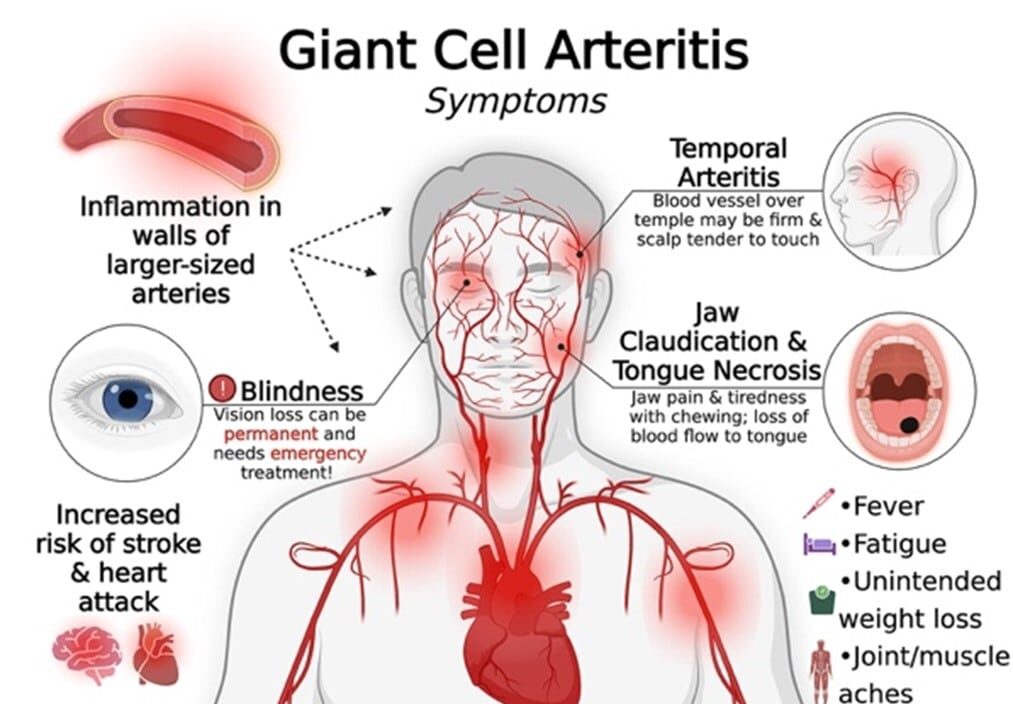
Imagine waking with a severe headache worse than any you have ever experienced. Your vision blurs. Suddenly, your sight in one eye disappears completely. Minutes later, your other eye goes dark. Within hours, you are permanently blind. This catastrophic scenario is the devastating reality for patients with untreated giant cell arteritis—a serious autoimmune condition causing inflammation of blood vessels, particularly in the head. Vision loss from giant cell arteritis is usually painless, sudden, and permanent. Without immediate treatment, blindness develops in up to 50 percent of untreated patients. However, with prompt recognition and aggressive treatment, blindness can usually be prevented. This stark contrast between treated and untreated disease makes recognizing giant cell arteritis early crucial for preventing devastating disability. Giant cell arteritis, also called temporal arteritis, is a chronic autoimmune inflammatory disease affecting medium and large blood vessels, particularly temporal arteries in the temples. The inflammation causes swelling and narrowing of arteries, reducing blood flow to tissues supplied by those arteries. If temporal arteries narrow severely, blood flow to the eyes decreases. This can cause sudden, permanent blindness—the most feared complication. Other arteries including the aorta, carotid arteries, and arteries to the limbs can also be affected, causing other serious complications. Giant cell arteritis almost exclusively affects people over age 50, with most cases occurring in people over 70. Women are more likely than men to develop the disease. The disease is closely related to polymyalgia rheumatica—approximately 50 percent of giant cell arteritis patients also have polymyalgia rheumatica symptoms. Approximately 1 in 500 people over age 70 develop giant cell arteritis. Despite relative rarity, the seriousness of potential vision loss makes recognition and prompt treatment crucial. Modern treatment with corticosteroids is highly effective. High-dose corticosteroids rapidly reduce inflammation preventing vision loss. However, treatment must begin immediately upon symptom recognition—delays can lead to irreversible blindness. In this comprehensive article, we will explore what giant cell arteritis is, understand how blood vessel inflammation develops, recognize warning signs requiring urgent evaluation, learn about serious complications, understand how diagnosis is made, explore treatments that save vision, and discover management strategies for this serious condition.
Understanding Blood Vessels and Arteritis
Before we explore giant cell arteritis, we need to understand blood vessels and what happens when they become inflamed. Blood vessels are hollow tubes transporting blood throughout your body. Arteries carry oxygen-rich blood from the heart to tissues. Veins return blood to the heart. Arteries have multiple layers. The innermost layer is the intima. The middle layer is the media, composed of smooth muscle allowing the artery to change diameter. The outermost layer is the adventitia, providing structural support. Different arteries have different thicknesses—large arteries like the aorta have thick walls, while small arteries have thin walls. Blood vessel inflammation is a normal response to injury or infection. When blood vessels are injured or infected, inflammation increases—immune cells travel to the area producing inflammatory chemicals causing swelling. This inflammation brings healing factors promoting repair. The swelling causes arteries to narrow, reducing blood flow. The reduced blood flow can cause symptoms—pain, weakness, or loss of function in tissues supplied by the narrowed artery. In giant cell arteritis, the immune system mistakenly attacks artery walls. Inflammation becomes chronic and inappropriate—the immune system continuously attacks healthy artery tissue. T lymphocytes and other immune cells infiltrate artery walls producing inflammatory chemicals. The inflammation causes artery wall swelling and thickening. The swelling narrows the artery opening, reducing blood flow. If the narrowing is severe, blood flow stops completely. Tissues supplied by the blocked artery become oxygen-deprived. If this happens in temporal arteries supplying the eye, the retina becomes oxygen-deprived and dies. Retinal death causes permanent vision loss—blindness that cannot be reversed. The temporal arteries are particularly important because they supply blood to the eyes. The ophthalmic artery branches from the internal carotid artery and supplies the retina through the central retinal artery. If giant cell arteritis narrows these arteries, vision is threatened. Inflammation can also affect other arteries including the aorta, causing aortitis. Aortitis can weaken the aorta causing aneurysm—bulging and potential rupture, which is life-threatening. The carotid arteries supplying the brain can be affected, increasing stroke risk. Understanding that giant cell arteritis involves inflammation causing artery narrowing and reduced blood flow helps explain the symptoms and why prompt treatment preventing further inflammation is crucial.
What is Giant Cell Arteritis?
Giant cell arteritis is a chronic autoimmune inflammatory disease affecting medium and large blood vessels, causing inflammation, artery wall thickening, and blood vessel narrowing. The disease is called “giant cell” arteritis because microscopic examination of affected arteries shows characteristic giant cells—large cells formed from fused immune cells. Giant cells help wall off inflammation but also cause artery destruction. Giant cell arteritis is also called temporal arteritis because temporal arteries in the temples are frequently affected. However, the disease can affect other arteries including the aorta, carotid arteries, and arteries to the arms and legs. The disease is also called polymyalgia rheumatica with arteritis when both conditions are present. Giant cell arteritis is fundamentally an autoimmune disease—the body’s immune system mistakenly attacks the body’s own artery walls. T lymphocytes and macrophages infiltrate artery walls producing inflammatory chemicals. The inflammatory response damages artery walls and causes swelling. What causes the immune system to attack artery walls in giant cell arteritis is not completely understood. Genetic factors are important—the disease runs in families. Specific genetic markers increase susceptibility. However, genetics alone does not cause giant cell arteritis—environmental factors are also necessary. Infections have been suspected as potential triggers. Some viral infections might trigger autoimmune response in genetically predisposed individuals. Age is the strongest risk factor—giant cell arteritis almost exclusively affects people over 50, with most cases in people over 70. The reason why autoimmune arteritis develops predominantly in older age is unclear. Some researchers suggest aging immune systems make arteritis development more likely. Others suggest lifetime environmental exposures accumulate triggering disease in later life. Giant cell arteritis develops suddenly in people who were previously healthy. The disease causes significant morbidity—blood vessel inflammation causes serious complications including blindness, stroke, and heart attack. However, giant cell arteritis responds dramatically to corticosteroid treatment. High-dose corticosteroids rapidly reduce inflammation preventing vision loss and other serious complications. This dramatic response to corticosteroids is one of the most striking features of the disease.
How Blood Vessel Inflammation Causes Vision Loss
Understanding how giant cell arteritis causes vision loss helps explain why this disease is so serious and why immediate treatment is crucial. Vision loss from giant cell arteritis occurs through artery narrowing reducing blood flow to the eye. The temporal arteries in the temples supply blood to the eye through the ophthalmic artery, which branches to the central retinal artery supplying the retina. The retina is the light-sensitive tissue at the back of the eye. When light hits the retina, specialized cells called photoreceptors convert light into electrical signals transmitted to the brain through the optic nerve. The brain interprets these signals as vision. The retina requires constant blood supply delivering oxygen and nutrients. If blood supply is cut off, retinal cells die within minutes. Dead retinal cells cannot regenerate. Vision loss from retinal death is permanent. In giant cell arteritis, inflammation of temporal arteries causes swelling narrowing the artery opening. If inflammation is severe, the artery narrows dramatically. Blood flow through the narrowed artery decreases. The eye becomes oxygen-deprived. If blood flow stops completely, the retina becomes oxygen-deprived. Retinal cells die within minutes. The person experiences sudden vision loss. The vision loss is painless—the eye does not hurt. The person simply notices vision disappearing. This painless vision loss is characteristic of arteritic vision loss and distinguishes it from other causes of vision loss which might cause eye pain. Anterior ischemic optic neuropathy (AION) is the most common mechanism of vision loss in giant cell arteritis. The optic nerve head—where the optic nerve enters the eye—becomes oxygen-deprived. The optic nerve cells die. Vision loss from optic nerve death affects specific parts of the visual field—classically, vision loss is altitudinal, affecting the upper or lower half of the visual field. Central retinal artery occlusion (CRAO) occurs when the central retinal artery completely blocks. This causes sudden, complete blindness in the affected eye. Amaurosis fugax is transient vision loss lasting minutes. Vision returns to normal after vision loss resolves. This is a warning sign that permanent vision loss might be imminent. Amaurosis fugax indicates critical artery narrowing. It is a medical emergency warning that immediate treatment is necessary. Posterior ciliary artery occlusion affects arteries supplying the back of the eye. This causes vision loss affecting specific visual field areas. Multiple arteries can be affected in the same eye or different eyes, causing various vision loss patterns. The key to preventing permanent vision loss is recognizing warning signs like amaurosis fugax and initiating high-dose corticosteroid treatment immediately. If corticosteroids are started before retinal death occurs, the inflammation decreases preventing further vision loss. However, if vision is already lost before treatment begins, the lost vision cannot be restored. This is why immediate medical attention upon any vision change is crucial.
Early Warning Symptoms: Recognizing the Emergency
Giant cell arteritis causes symptoms that should prompt immediate medical evaluation. Recognizing these warning signs and seeking urgent care can prevent permanent vision loss. Severe headache is one of the most common early symptoms. The headache is often the worst headache the person has ever experienced. The headache is typically localized to the temples but can be diffuse. The headache is often resistant to usual pain medications. Severe, new-onset headache in someone over 50 is a red flag requiring urgent evaluation. Scalp tenderness is extremely common. The scalp becomes tender when touched. Even combing hair causes discomfort. Some patients describe the scalp feeling bruised. Scalp tenderness is often localized to the temples where temporal arteries are most prominent. Vision changes are the most serious warning sign. Blurred vision, double vision, or dimming of vision requires urgent evaluation. Transient vision loss (amaurosis fugax) lasting minutes is a medical emergency—it indicates critical artery narrowing and is often followed by permanent vision loss if untreated. Sudden complete vision loss in one eye is a medical emergency. This indicates central retinal artery occlusion. Immediate high-dose corticosteroid treatment can sometimes prevent vision loss in the other eye. Jaw claudication is a distinctive symptom. Jaw pain develops during chewing. Patients must stop eating due to pain. Jaw claudication is caused by ischemia in jaw muscles from reduced blood supply. This symptom strongly suggests giant cell arteritis. Arm claudication—pain in arms during activity—indicates arteritis affecting arm arteries. Pain develops with activity and resolves with rest. Leg claudication similarly indicates arteritis of leg arteries. Fever is common. Low-grade fever around 100 to 101 degrees Fahrenheit is typical. Fatigue is nearly universal. Patients report overwhelming exhaustion. Weight loss occurs in some patients. These constitutional symptoms indicate systemic inflammation. Myalgia (muscle pain) and arthralgia (joint pain) occur in about 50 percent of giant cell arteritis patients. Patients might have polymyalgia rheumatica symptoms—shoulder and hip pain and stiffness. These symptoms help recognize the giant cell arteritis/polymyalgia rheumatica connection. Transient neurologic symptoms including TIA-like symptoms (temporary weakness, speech difficulties) might indicate early stroke from arteritis affecting cerebral arteries. These symptoms require urgent evaluation. Any new symptoms in someone over 50 with risk factors for giant cell arteritis warrant prompt medical evaluation. Vision changes, headache, scalp tenderness, or jaw pain are particularly concerning and require emergency evaluation.
How Doctors Diagnose Giant Cell Arteritis
Diagnosing giant cell arteritis requires combining clinical findings, blood tests, and sometimes imaging or biopsy studies. The diagnosis must be made quickly because vision-saving treatment must begin immediately. Clinical history is crucial. Doctors ask about symptom onset—sudden onset of severe headache, vision changes, or scalp tenderness in someone over 50 is classic. They ask about constitutional symptoms including fever, fatigue, and weight loss. They ask about polymyalgia rheumatica symptoms. Physical examination assesses vital signs and carefully examines the head and arteries. Blood pressure is typically elevated. Temporal arteries are palpated—inflamed arteries might be tender or prominent. Cranial nerves are examined, particularly vision and eye movements. Blood tests are performed immediately. Inflammatory markers including erythrocyte sedimentation rate (ESR) and C-reactive protein (CRP) are typically markedly elevated. In giant cell arteritis, ESR is often over 80 mm/hour (normal is under 20). CRP is elevated. However, about 5 to 10 percent of giant cell arteritis patients have normal inflammatory markers despite classic symptoms. Complete blood count sometimes shows anemia. Hemoglobin is sometimes low from chronic inflammation. Platelet elevation sometimes occurs. Blood chemistry assesses kidney and liver function. Temporal artery biopsy is the gold standard diagnostic test. A small artery sample is obtained from the temple area. Microscopic examination shows characteristic findings of giant cell arteritis including giant cells, lymphocytic infiltration, and artery wall thickening. A positive biopsy confirms diagnosis. However, biopsy is not always necessary if clinical and blood test findings are highly suggestive. Some patients have negative biopsy despite clinical giant cell arteritis—this occurs because inflammation is patchy, and the biopsy might sample normal artery. Temporal artery ultrasound shows arterial wall thickening characteristic of giant cell arteritis. Ultrasound can be useful in diagnosing arteritis. Ultrasound is non-invasive and might reduce need for biopsy. Color doppler ultrasound shows reduced blood flow through narrowed arteries. PET scan or CT angiography can show inflammation throughout the body in patients with systemic arteritis. These imaging studies are useful when aortitis or other arteritis is suspected. Immediate ophthalmologic evaluation is crucial if vision changes have occurred. Ophthalmologists examine the retina and optic nerve assessing for ischemic changes. Visual field testing might show areas of vision loss. OCT imaging shows optic nerve changes. Platelet count elevation might occur. Liver enzyme elevation sometimes occurs. These laboratory findings support diagnosis. The key to good outcomes is rapid diagnosis and immediate treatment initiation. Waiting for confirmatory biopsy results while vision is threatened is inappropriate. High-dose corticosteroid treatment should begin immediately when giant cell arteritis is suspected based on clinical and blood test findings. Biopsy can be performed while corticosteroids are already being given.
Treatment: Vision-Saving Therapy
Giant cell arteritis treatment aims to rapidly reduce inflammation preventing vision loss and other serious complications. Treatment must begin immediately upon diagnosis suspicion. High-dose corticosteroids are the standard treatment. Prednisone at very high doses—typically 40 to 60 mg daily—is started immediately. Some patients with vision-threatening disease receive even higher doses or intravenous methylprednisolone. High-dose corticosteroids rapidly reduce inflammation. Within 24 to 48 hours, inflammatory markers decrease significantly. Symptoms improve dramatically. If vision is threatened, immediate high-dose corticosteroids can sometimes prevent vision loss. Patients are monitored closely during initial treatment. Within days, symptoms including headache, scalp tenderness, and jaw claudication improve. Fever and fatigue resolve. If vision loss has not occurred before treatment, corticosteroids usually prevent it. The dramatic response to corticosteroids within hours to days is one of the most striking features of giant cell arteritis. Once remission is achieved, corticosteroid doses are gradually tapered. Doses are reduced slowly over months. Too-rapid tapering causes disease flares. Many patients require ongoing low-dose corticosteroids (5 to 10 mg prednisone daily) for months or years. Inflammatory markers are monitored during taper—if markers increase suggesting disease activity, dose reduction is paused or reversed. Immunosuppressive medications including methotrexate are sometimes used to reduce corticosteroid requirements. These medications suppress immune activity complementing corticosteroid effect. Using methotrexate alongside corticosteroids allows lower corticosteroid doses in some patients. Tocilizumab, an IL-6 inhibitor, shows promise for giant cell arteritis. This biologic medication blocks IL-6, a key inflammatory chemical. Tocilizumab might reduce corticosteroid requirements and improve outcomes. Aspirin is sometimes recommended for patients with giant cell arteritis. Aspirin reduces clotting helping prevent blood vessel closure. However, evidence for aspirin benefit is mixed. Calcium and vitamin D supplementation is crucial. Corticosteroids increase bone loss. Calcium and vitamin D supplementation helps prevent osteoporosis. Bone density monitoring assesses for osteoporosis development. Bone density medication might be prescribed if osteoporosis develops. Management of corticosteroid side effects is necessary. Weight gain, mood changes, and sleep disturbances are common. Dietary modifications help manage weight. Blood pressure monitoring is important—corticosteroids increase blood pressure. Regular monitoring and medication adjustment prevents hypertension.
Living with Giant Cell Arteritis: Long-term Management
Living with giant cell arteritis requires ongoing corticosteroid management, monitoring for complications, maintaining treatment adherence, and psychological adaptation. Taking corticosteroids exactly as prescribed is absolutely essential. Missing doses allows inflammation to rebound and vision loss can recur. Regular dosing maintains inflammation suppression. Gradual tapering under doctor supervision allows eventual discontinuation or minimum maintenance dose. Attending rheumatology appointments regularly ensures disease monitoring. Regular visits assess disease activity and corticosteroid response. Blood tests monitor inflammatory markers and medication side effects. Adjustments to corticosteroid doses are made based on symptoms and marker levels. Monitoring for vision problems is crucial. Any new vision symptoms require immediate evaluation. Regular eye exams help monitor vision and detect complications early. Visual field testing assesses for early vision changes. Immediate reporting of any vision symptoms to eye doctors might allow intervention preventing vision loss. Monitoring for other arteritis complications is important. Watching for new headaches, scalp tenderness, or jaw pain might indicate disease flares requiring treatment adjustments. Watching for arm or leg pain during activity indicates possible peripheral artery involvement. Watching for stroke symptoms including weakness or speech changes indicates possible cerebral artery involvement. Regular blood pressure monitoring helps detect hypertension from corticosteroids or arteritis. Blood pressure medication might be necessary. Bone density monitoring assesses osteoporosis risk. Bone density medication might be prescribed if osteoporosis develops. Calcium and vitamin D supplementation is important. Adequate calcium and vitamin D intake helps prevent bone loss. Regular physical activity helps maintain strength and bone density. Gentle exercise is usually well-tolerated. Walking, swimming, or gentle yoga help maintain fitness. Mental health support helps cope with chronic disease. Depression is common with chronic illness and corticosteroid use. Counseling or antidepressants help some patients. Accepting disease and adapting to limitations helps psychological adjustment. Stress management helps. Relaxation techniques, meditation, or counseling help manage stress. Pursuing enjoyable activities brings psychological benefit. Sleep optimization is important. Sleep deprivation can worsen symptoms. Good sleep habits help. Corticosteroid-induced insomnia might require medication. Nutrition supports healing and bone health. Adequate calories and protein support overall health. Adequate calcium and vitamin D intake prevents bone loss. Regular dental care maintains oral health—corticosteroids increase infection risk. Regular dental visits and good oral hygiene prevent problems. Eye care is paramount. Regular eye exams monitoring vision are crucial. Immediate reporting of any vision changes allows prompt evaluation. Using protective eyewear in bright sun helps prevent light-related eye discomfort. Family and social support is invaluable. Educating loved ones about giant cell arteritis helps them understand the seriousness and need for adherence. Support groups provide understanding from others managing similar conditions. Open communication helps relationships navigate disease impact.
Frequently Asked Questions (FAQs)
Q1: Can someone regain vision lost to giant cell arteritis?
Unfortunately, vision loss from giant cell arteritis is usually permanent. Once retinal cells or optic nerve cells die from lack of blood supply, they cannot regenerate. The lost vision cannot be restored with current medical treatments. This is why prevention is so crucial—preventing vision loss is far easier than restoring it after it occurs. With immediate high-dose corticosteroid treatment before vision loss develops, blindness can usually be prevented. However, if vision has already been lost before treatment begins, that vision loss is generally permanent.
Q2: Why is immediate treatment so important in giant cell arteritis?
Immediate treatment is crucial because vision loss from giant cell arteritis can develop within hours to days of symptom onset. Without immediate high-dose corticosteroid treatment, vision loss risk is very high. With immediate treatment, vision can usually be preserved. The window for preventing vision loss is narrow—days at most. Delayed diagnosis or delayed treatment initiation dramatically increases risk of permanent blindness. This is why any new symptoms including headache, vision changes, or scalp tenderness in someone over 50 warrant immediate evaluation. Any vision change requires emergency evaluation.
Q3: Is giant cell arteritis curable?
Giant cell arteritis cannot be cured with current treatments. The underlying autoimmune dysfunction is permanent. However, with appropriate corticosteroid treatment, inflammation can be suppressed and remission can be achieved. Most patients eventually discontinue corticosteroids after months or years. Some require ongoing low-dose corticosteroids indefinitely. With proper management and monitoring for complications, most giant cell arteritis patients avoid serious complications and maintain quality of life. The key is early diagnosis and prompt treatment.
Q4: What happens if giant cell arteritis is not treated?
Untreated giant cell arteritis causes serious complications. Vision loss from arteritic anterior ischemic optic neuropathy or central retinal artery occlusion develops in up to 50 percent of untreated patients. Blindness is permanent. Stroke from arteritis of cerebral arteries causes neurologic disability. Myocardial infarction from coronary artery involvement can be fatal. Aortic aneurysm from aortitis can rupture causing death. These serious complications emphasize the absolute necessity of immediate diagnosis and treatment.
Q5: Can giant cell arteritis recur after treatment ends?
Giant cell arteritis can recur if corticosteroids are discontinued. Disease flares sometimes occur during corticosteroid tapering. If inflammatory markers increase or symptoms recur, corticosteroid doses must be increased. Some patients experience multiple flares requiring prolonged treatment. Others achieve sustained remission after discontinuing corticosteroids. Recurrence is more common if corticosteroid tapering is too rapid. Careful, gradual tapering under medical supervision minimizes flare risk. Some patients require ongoing low-dose corticosteroids indefinitely to prevent recurrence.
Key Takeaways
Giant cell arteritis is a chronic autoimmune inflammatory disease affecting blood vessels, particularly temporal arteries in the temples. The disease almost exclusively affects people over age 50, with most cases occurring after age 70. Inflammation of temporal arteries causes artery narrowing reducing blood flow to the eye. Severe narrowing can cause sudden, permanent blindness—the most feared complication. Approximately 50 percent of untreated giant cell arteritis patients develop vision loss. Early warning signs including severe headache, scalp tenderness, vision changes, and jaw claudication require immediate medical evaluation. Transient vision loss (amaurosis fugax) is a medical emergency indicating critical artery narrowing and imminent risk of permanent blindness. Diagnosis combines clinical findings, elevated inflammatory markers (ESR and CRP), and temporal artery biopsy showing giant cells. High-dose corticosteroids started immediately can prevent vision loss and other serious complications. Vision loss prevented before it occurs can usually be avoided with prompt treatment. Approximately 50 percent of giant cell arteritis patients also have polymyalgia rheumatica symptoms. Corticosteroids are gradually tapered over months but many patients require ongoing therapy. Long-term complications including aortitis, stroke, and heart attack can occur. With proper diagnosis and immediate treatment, most giant cell arteritis patients avoid vision loss and maintain good quality of life. Early recognition is literally sight-saving.
References
- World Health Organization (WHO). “Giant Cell Arteritis and Temporal Arteritis.” Retrieved from https://www.who.int/
- American College of Rheumatology. “Giant Cell Arteritis: Clinical Guidelines and Resources.” Retrieved from https://www.rheumatology.org/
- Mayo Clinic. “Giant Cell Arteritis: Causes, Symptoms, and Treatment.” Retrieved from https://www.mayoclinic.org/
- Cleveland Clinic. “Giant Cell Arteritis: Complete Information and Management.” Retrieved from https://my.clevelandclinic.org/
- American Academy of Ophthalmology. “Giant Cell Arteritis and Vision Loss Prevention.” Retrieved from https://www.aao.org/
- National Eye Institute. “Giant Cell Arteritis Information.” Retrieved from https://www.nei.nih.gov/
Related Articles on ObserverVoice.com
Explore more health and science topics on our platform:
- Polymyalgia Rheumatica: The Older Adult Disease Behind Unexplained Muscle Stiffness
- Understanding Autoimmune Diseases: When Your Immune System Attacks Itself
- The Cardiovascular System: Heart and Blood Vessels Health
- Vision Health: Protecting Your Eyes from Disease
- Aging and Health: Common Conditions Affecting Older Adults
- Living with Chronic Illness: Adaptation and Support Strategies
Disclaimer
This article adapts publicly available information from WHO’s Giant Cell Arteritis and Temporal Arteritis page. This content is for informational and educational purposes only and does not constitute medical advice. [ObserverVoice.com] is a news and information platform — not a healthcare provider. If you suspect you have giant cell arteritis, experiencing severe headache, vision changes, scalp tenderness, or jaw pain, seek immediate medical attention. These symptoms constitute a medical emergency—vision-threatening arteritis requires urgent evaluation and treatment to prevent permanent blindness. Do not delay seeking emergency care. Any new vision symptoms including blurred vision, vision loss, or transient vision loss require immediate ophthalmologic evaluation. Call 911 or go to the nearest emergency room if experiencing acute vision loss. Early diagnosis and immediate high-dose corticosteroid treatment prevent serious complications including permanent blindness. Always seek guidance from licensed rheumatologists and ophthalmologists for proper diagnosis and management of giant cell arteritis.
Observer Voice is the one stop site for National, International news, Sports, Editor’s Choice, Art/culture contents, Quotes and much more. We also cover historical contents. Historical contents includes World History, Indian History, and what happened today. The website also covers Entertainment across the India and World.

